
South Asia Pvt. Ltd. Technical Field Report

South Asia Pvt. Ltd. Technical Field Report
Pharmacological Basis, Clinical Applications, and Practical Considerations for Drinking-Water Administration
The Economic Impact of Bacterial Diseases in Poultry
Bacterial infections continue to impose significant economic losses throughout the poultry value chain. Among the most important bacterial disease syndromes encountered in broiler production are:
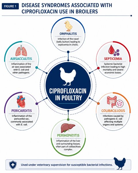
Escherichia coli remains one of the most frequently isolated bacterial pathogens associated with these conditions. The first week of life is particularly critical, as early chick mortality associated with bacterial infections can have long-term consequences on flock performance.
Disease Syndromes Associated with Ciprofloxacin Use in Broilers.
Why Rapid Intervention Matters
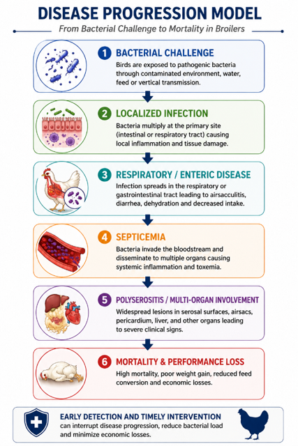
The progression of bacterial infections in poultry can be remarkably rapid.
What begins as localized bacterial colonization may quickly develop into respiratory involvement, systemic septicemia, polyserositis, and ultimately mortality. As disease progresses, treatment becomes increasingly difficult and production losses become more severe.
The success of antimicrobial therapy often depends not only on antimicrobial selection but also on the speed with which effective treatment is initiated.
Ciprofloxacin as a Solution:
The ideal antimicrobial for modern poultry production should possess:
• Rapid bactericidal activity • Broad-spectrum efficacy against susceptible bacterial pathogens
• Excellent tissue penetration • Systemic distribution • Practical administration characteristics
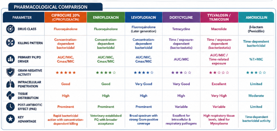
Ciprofloxacin possesses many of these attributes. As a second-generation fluoroquinolone, ciprofloxacin exerts its antibacterial activity through inhibition of bacterial DNA gyrase and topoisomerase IV, resulting in concentration-dependent bacterial killing.
Unlike bacteriostatic agents that inhibit bacterial growth, ciprofloxacin rapidly reduces bacterial populations when adequate tissue concentrations are achieved.
Pharmacokinetic studies have demonstrated favorable oral absorption, broad tissue distribution, and effective systemic exposure following administration in broiler chickens.
These characteristics make ciprofloxacin particularly suitable for bacterial infections involving respiratory tissues, serosal surfaces, and internal organs.
Clinical Applications in Broiler Production
Although ciprofloxacin is often associated with colibacillosis, its clinical utility extends beyond a single disease condition.
Its use may be considered in susceptible bacterial infections associated with:
• Colibacillosis • Colisepticemia • Omphalitis • Yolk sac infections • Airsacculitis
• Pericarditis • Perihepatitis • Secondary bacterial respiratory infections
• Septicemic disease complexes
The broad tissue distribution achieved following oral administration allows ciprofloxacin to reach multiple target sites commonly affected during systemic bacterial infections.
Why Route of Administration Influences Treatment Success
“In modern broiler production, therapeutic success depends not only on antimicrobial efficacy but also on the speed, uniformity, and reliability of drug delivery.”
Experimental Evidence Supporting Clinical Use of Ciprofloxacin: Several studies have demonstrated the effectiveness of ciprofloxacin in controlling susceptible bacterial infections.
In an experimental E. coli challenge model, ciprofloxacin-treated broilers demonstrated significantly lower mortality, reduced bacterial recovery, improved clinical recovery, and superior production performance compared with untreated challenged birds.
Mortality Reduction in Experimental E. coli Challenge.
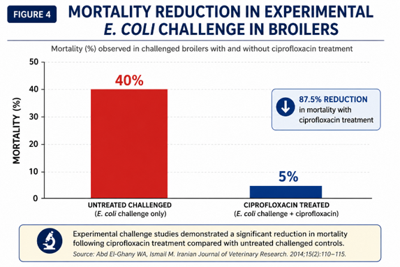
These findings reinforce the importance of rapid intervention and effective antimicrobial therapy during bacterial disease outbreaks.
Conclusion
Modern poultry production requires both effective antimicrobial therapy and efficient delivery systems to successfully manage bacterial disease challenges.
Ciprofloxacin combines concentration-dependent bactericidal activity, favorable pharmacokinetic characteristics, broad tissue distribution, and suitability for drinking-water administration, making it an important therapeutic option in the management of susceptible bacterial infections in broilers.
When combined with rapid diagnosis, appropriate administration, and responsible antimicrobial stewardship, ciprofloxacin continues to play a valuable role in contemporary poultry health management.
Furthermore, oral liquid formulations can enhance the practicality of water medication programs through improved ease of handling, rapid dispersion, and greater operational convenience under commercial production conditions.
Dr. Kishor Gedam, Dr. Sanjay Singhal
Abstract
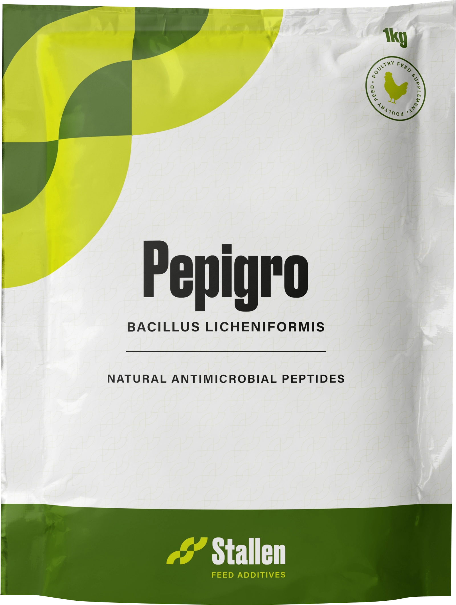
This study evaluates the efficacy of PEPIGRO, a Bacillus licheniformis-based probiotic and postbiotics as antimicrobial peptide (AMPs), on the growth and health performance of commercial broilers under field conditions. A total of 36,000 straight-run broiler chicks were assigned to control and treatment groups, with the latter receiving PEPIGRO supplementation at 300 g/ton of feed. The trial was conducted over 42 days during extreme heat (42–45°C), and assessed body weight, feed intake, feed conversion ratio (FCR), weekly gain, and mortality. PEPIGRO supplementation resulted in an 8.18% increase in body weight, a 6.59% rise in feed intake, and a 6.22% improvement in weekly gain compared to the control, alongside a 1.68% enhancement in FCR. Mortality was notably reduced by 28.08%, indicating improved survivability. These findings demonstrate that dietary inclusion of PEPIGRO effectively enhances broiler growth performance, feed efficiency, and health, supporting the role of Bacillus licheniformis as a promising antibiotic alternative under commercial field stressors.
1. Introduction
The widespread use of antibiotics in animal husbandry for growth promotion and disease control has led to serious concerns, including antibiotic resistance and environmental contamination (Tang et al., 2017). Consequently, restrictions on antibiotic growth promoters (Organization, 1999) have accelerated the search for safer alternatives. Among these, bioactive feed additives, such as probiotics, antimicrobial peptides, plant extracts, acidifiers, and essential oils—have shown potential in improving growth, immunity, oxidative balance, and gut health (Xu et al., 2021; Yi et al., 2017; Pearlin et al., 2020; Montassier et al., 2021).
Probiotics, particularly Bacillus licheniformis, have gained attention due to their safety and multifunctional benefits (Ningsih et al., 2023). This spore-forming bacterium enhances nutrient digestion through enzyme production, modulates gut microbiota, suppresses pathogens, and improves immune responses (Giri et al., 2019). It also produces antimicrobial compounds and enhances antioxidant activity, contributing to better intestinal integrity and performance (Jia et al., 2018; Chen and Yu, 2020).
Necrotic enteritis (NE), caused by Clostridium perfringens, is a major poultry disease causing significant economic losses (Wade and Keyburn, 2015). Probiotics like B. licheniformis have demonstrated potential in mitigating NE by improving gut barrier function, modulating immunity, and stabilizing microbiota (Wang et al., 2017; Lin et al., 2017).
2. Antimicrobial Peptides (AMPs)
Antimicrobial peptides (AMPs) are small, naturally occurring bioactive molecules found in diverse organisms and play a key role in innate immunity as a first line of defense. They exhibit broad-spectrum activity against bacteria, fungi, parasites, and viruses, contributing significantly to host protection (Huan et al., 2020).
Antibacterial Substances Produced by Bacillus licheniformis
The endospore-forming bacterium Bacillus licheniformis produces a wide range of antimicrobial compounds with diverse structural and functional properties, typically ranging from 1.4 to 20 kDa. These include bacteriocins, licheniformins, bacitracin, and surfactin (Shleeva et al., 2023).
2.1. Bacteriocins
Bacteriocins are ribosomally synthesized antimicrobial peptides or proteins that exhibit bactericidal or bacteriostatic activity against closely related bacteria. Bacillus licheniformis produces various bacteriocins (1.4–55 kDa), influenced by environmental conditions, growth phase, and strain genotype. For example, strain B116 secretes a ~4 kDa bacteriocin active against both Gram-positive and Gram-negative bacteria, including Staphylococcus aureus, Escherichia coli, and Salmonella spp. This compound is heat- and pH-resistant but is inactivated by pronase and partially affected by papain and lipase, suggesting a lipid component (Shleeva et al., 2023).
2.2. Licheniformins
Licheniformins are lipopeptide antibiotics produced by Bacillus licheniformis, often occurring as closely related variants. The licheniformin from strain MS3 has a molecular mass of ~1.438 kDa, while the main forms—licheniformins A, B, and C—range from 3.8 to 4.8 kDa with similar amino acid compositions. Despite structural similarity, they differ in antibacterial potency and toxicity due to variations in side chains and lipid modifications (Shleeva et al., 2023).
2.3. Bacitracin
Bacitracin is a well-known polypeptide antibiotic non-ribosomally synthesized by certain strains of B. subtilis and B. licheniformis. It is composed of 12 amino acids, with four of them—glutamic acid, aspartic acid, phenylalanine, and ornithine—present in their D-isomer forms. The molecular mass of bacitracin is approximately 1.42 kDa. Bacitracin functions by interfering with bacterial cell wall synthesis, making it a clinically important peptide used to inhibit Gram-positive pathogens (Shleeva et.al.2023).
2.4. Surfactin
Bacillus licheniformis produces cyclic lipopeptides such as surfactin and its analog lichenysin, known for strong surface-active and antimicrobial properties. Strain HSN221 secretes nine variants of these compounds under optimal culture conditions (glucose, ammonium chloride, and yeast extract). The surfactin monomethyl ester homologues have molecular masses of ~1.048–1.063 kDa (ESI-MS) and exhibit potent antimicrobial and emulsifying activities with applications in pharmaceutical, agricultural, and environmental biotechnology (Shleeva et al., 2023).
3.Mechanism Of Action
Bacillus linchiniformis promote gut health through complementary competitive and immunological mechanisms. First, they competitively exclude pathogens by adhering to intestinal mucosa, thereby occupying ecological niches and preventing pathogen attachment and invasion. They also compete for nutrients by secreting extracellular enzymes that efficiently utilize available macro- and micronutrients, limiting resources required for pathogenic growth. In addition, Bacillus produces antimicrobial metabolites, including lipopeptides, bacteriocins, polyketides, and short-chain fatty acids (SCFAs), which directly inhibit pathogenic microorganisms. Furthermore, oxygen consumption by Bacillus reduces intestinal oxygen levels, creating a favorable hypoxic environment for beneficial anaerobic and fermentative bacteria such as lactic acid bacteria.
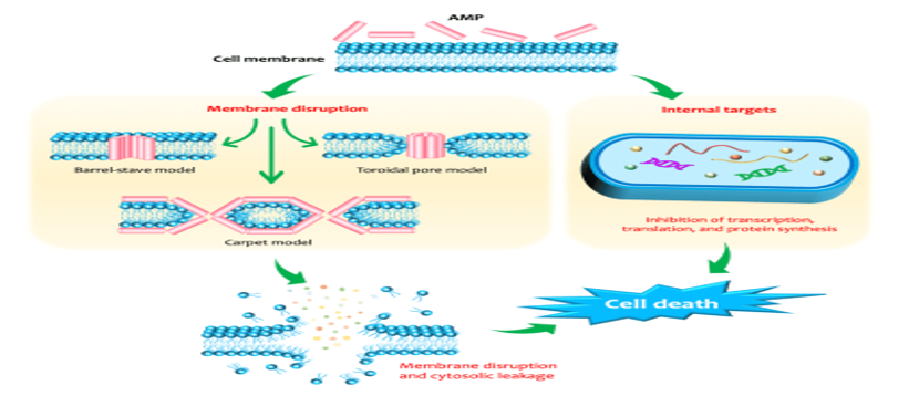
Simultaneously, antimicrobial peptides (AMPs) contribute to host defense through direct antimicrobial and immunomodulatory activities. AMPs regulate cytokine and chemokine production and modulate immune cells, including macrophages, dendritic cells, and lymphocytes, maintaining immune homeostasis. Mechanistically, AMPs disrupt microbial membranes via barrel-stave, carpet, or toroidal pore-forming models, leading to membrane destabilization and lysis. Additionally, they can penetrate cells and inhibit intracellular processes such as nucleic acid and protein synthesis, enzyme activity, and cell wall formation, thereby ensuring effective pathogen clearance and enhanced innate and adaptive immune responses.
Fig.1. Probiotic Bacillus employs multifactorial competition mechanism to restrict the expansion of pathogens through four pathways.
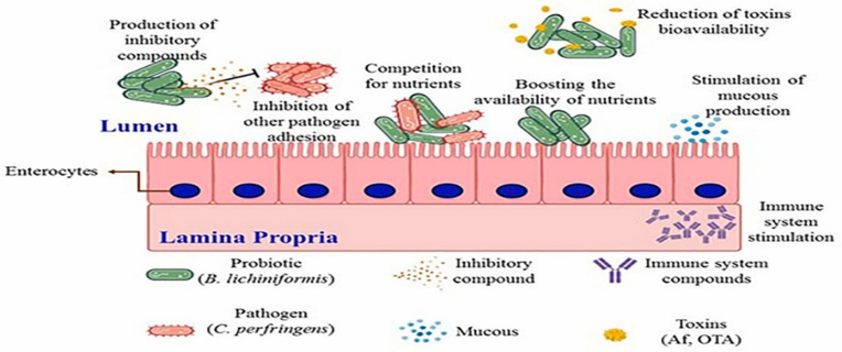
Fig.2. Models of antibacterial mechanisms of AMPs.
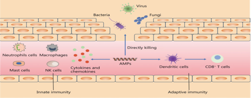
Fig.3. The membrane-disruptive and non-membrane-disruptive antibacterial mechanisms of antimicrobial peptides (AMPs).
4.MATERIALS AND METHODS
4.1. Experimental Design and Management
The trial was conducted at Harsh Broiler House, Bilaspur, using Vencobb 430 straight-run chicks (not sexed at the hatchery) in three treatments of around 12000 birds in each treatment. A total of 36000 birds were considered for trial purposes. The feed formulation used was the same for all treatment groups except in T3 where PEPIGRO (Bacillus licheniformis 3*109) was added at 300 gm per ton of feed, respectively, in all stages. (Table 1). In the study, the energy level was equivalent to the standard requirements of broilers recommended in the Vencobb 430. The trial was carried out over a period of 42 days. The birds were fed ad lib, and feed and water were available all the time. Care was taken to provide good conditions by adopting strict biosecurity measures. The housing and vaccination procedures were the same in both groups.
Table 1. Composition of basal diet for broiler chicks in control group for 3 phases.
| Broiler Feed Formulation (Control) | |||
| Raw Materials | Pre-starter | Starter | Finisher |
| Maize | 625.15 | 652.75 | 686.65 |
| HiPro Soya | 335 | 300 | 260 |
| Soya Crude Oil | 6 | 14 | 23 |
| Limestone Powder | 8.5 | 8.5 | 8 |
| Dicalcium Phosphate | 10 | 10 | 8 |
| L Lysine HCI | 2.7 | 2.4 | 2.3 |
| DL Methionine | 3.3 | 3 | 2.7 |
| L Threonine | 1 | 1 | 1 |
| Salt | 2.5 | 2.5 | 2.5 |
| Soda Bi Carb | 1.5 | 1.5 | 1.5 |
| Choline Chloride 60% | 1 | 1 | 1 |
| Organic TM | 0.5 | 0.5 | 0.5 |
| Broiler Vitamin Premix | 0.5 | 0.5 | 0.5 |
| Coccidiostat | 0.5 | 0.5 | 0.5 |
| AGP | 0.05 | 0.05 | 0.05 |
| NSP Enzyme | 0.1 | 0.1 | 0.1 |
| Phytase 5000 | 0.1 | 0.1 | 0.1 |
| Feed Acidifier | 1 | 1 | 1 |
| Toxin Binder | 0.6 | 0.6 | 0.6 |
*The figures are in Kilograms.
The premix provided the following per kilogram of the diet: vitamin A, 6000 IU; vitamin D3, 2500 IU; vitamin B1, 1.75 mg; vitamin B2, 5.5 mg; vitamin B6, 4 mg; vitamin B12, 0.18 mg; vitamin E, 25 mg; vitamin K3, 2.25 mg; Cu, 7.5 mg; Mn, 60 mg; Fe, 75 mg; Zn, 60 mg; Se, 0.15 mg; biotin, 0.14 mg; NaCl, 3.7 g; folic acid, 0.8 mg; pantothenic acid, 12 mg; phytase, 400 U; nicotinic acid, 34 mg; chloride, 350 mg. *Nutrient levels were all calculated values.
4.2. Treatment Details-
T1: Control group fed basal diet
T3: Control group fed basal diet + PEPIGRO @300 g PMT
4.3. Parameters Studied-
5.Result:
Effect of Pepigro on growth performance parameter in broiler.
Fig.4. Effect of different dietary treatments on Body Weights (g)
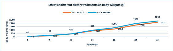
Conclusion: PEPIGRO supplementation at 300g/ton of feed (T3) resulted in a statistically significant 8.18% increase in broiler body weight compared to the control (T1), indicating improved growth performance.
Fig.5. Effect of different dietary treatment on Feed intake (g)
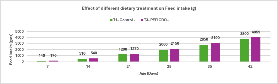
Conclusion: The broiler supplemented with PEPIGRO (T3) at 300g/ ton of feed had a feed intake of 4059 g, which is 6.59% higher than the control group (T1) with 3800 g feed intake. This increase in feed intake indicates that PEPIGRO supplementation positively influenced the birds’ feeding behaviour, likely by enhancing the palatability or nutrient availability of the diet.
Fig.6. Effect of different dietary treatment on Weekly Gain (g)
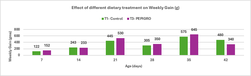
Conclusion: PEPIGRO (T3) supplementation in broiler diet at 300g/ton of feed resulted in the average percentage difference in weekly gain between T1 (Control) is approximately 6.22%. This indicates that PEPIGRO supplementation had a positive overall effect on growth performance, enhancing weight gain efficiency in broiler chickens.
Fig.7. Effect of different dietary treatment on Feed conversion ratio
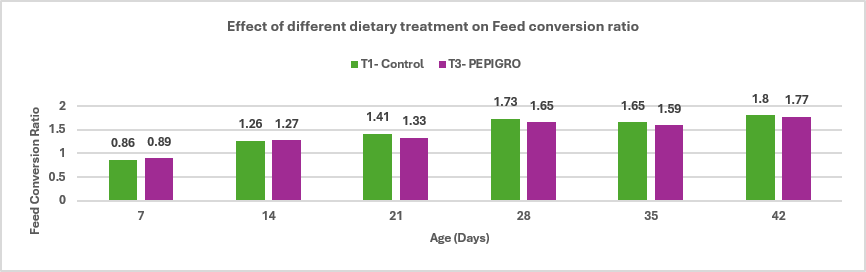
Conclusion: PEPIGRO (T3) supplementation in broiler diet at 300g/ton of feed resulted in a 1.68% improvement in feed conversion ratio (FCR) compared to the control group (T1), indicating enhanced feed efficiency and better growth performance.
Fig.8. Effect of different dietary treatment on Weekly mortality (%)
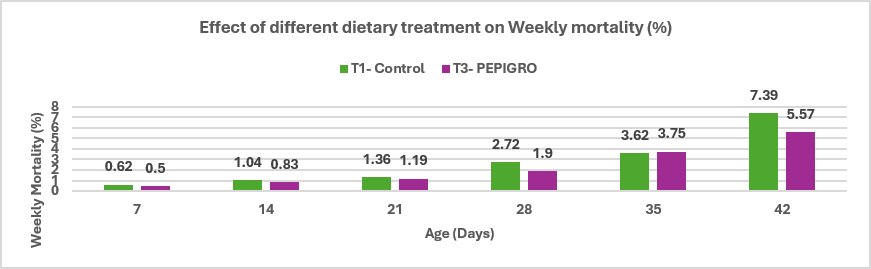
Conclusion: PEPIGRO supplementation at 300g/ton of feed reduced mortality in broiler poultry from 7.39% in the control group to 5.57%, reflecting a 28.08% decrease. This suggests that PEPIGRO may contribute to improved bird health and survivability during the rearing period.
Table 2. Summary of the Report
| Parameters | T1- Control | T3- PEPIGRO | % Difference |
| Body Weight (g) | 2110 | 2290 | 8.18 |
| Feed Intake (g) | 3800 | 4059 | 6.59 |
| FCR | 1.8 | 1.77 | 1.68 |
| CFCR | 1.77 | 1.69 | 4.62 |
| Mortality (%) | 7.39 | 5.57 | 28.08 |
6. Discussion
The discussion for this article highlights the significant positive effects of PEPIGRO, a Bacillus licheniformis-based probiotic, on the growth performance, feed efficiency, and health status of commercial broilers under field conditions. The 8.18% increase in body weight and 6.59% increase in feed intake, along with improvements in feed conversion ratio (FCR), align well with previous studies showing Bacillus probiotics enhance nutrient digestibility, modulate gut microbial populations, and improve intestinal morphology (Pan et al., 2022; Hung et al., 2019). These effects are especially valuable in the context of rising restrictions on antibiotic growth promoters (Tang et al., 2017), pushing for safer and sustainable alternatives.
The notable 28.08% reduction in mortality observed in this study suggests enhanced resilience of broilers to environmental stressors, likely owing to improved gut barrier integrity and immune modulation. Bacillus licheniformis produces antimicrobial peptides, enzymes, and metabolites such as bacteriocins and surfactins that inhibit pathogens like Clostridium perfringens, a major agent of necrotic enteritis (NE) in poultry (Shleeva et al., 2023; Wade and Keyburn, 2015). PEPIGRO’s capacity to maintain intestinal health and microbial balance may underlie the reduced pathogenic infections and inflammation, consistent with findings that show Bacillus supplementation upregulates tight junction proteins and mucins while enhancing beneficial microbes like Lactobacillus (Chen and Yu, 2020; Wang et al., 2017).
Moreover, the probiotic’s ability to stimulate the host immune system by inducing cytokine production and activating phagocytic cells further supports its protective role in the gut environment (Babakuliyev et al., 2022). This immunomodulatory effect is critical for mitigating subclinical infections and improving overall flock welfare, which translates into better productivity under commercial rearing conditions.
Additionally, PEPIGRO contributes to antioxidant status improvement by elevating enzyme activities such as superoxide dismutase and glutathione peroxidase, reducing oxidative stress that commonly compromises poultry health under heat stress conditions (Jia et al., 2018). This antioxidant benefit complements its antimicrobial and immunomodulatory functions.
In conclusion, this study reinforces the role of Bacillus licheniformis as a multifunctional probiotic that enhances growth performance, feed efficiency, and health in broilers. It offers a sustainable alternative to antibiotics, aligning with global efforts to reduce antibiotic use in animal production. Future studies should explore optimal dosing strategies, combinations with other feed additives, and long-term effects on microbiota composition and immune function to fully harness the benefits of PEPIGRO in commercial poultry systems.
7. Conclusion-
The trial was conducted in the extreme heat season where average temperature in the surrounding was around 42-45 degree Celsius. The T3 (PEPIGRO) group showed notable improvements compared to the T1 (Control) group. Body weight in T3 (PEPIGRO) increased by 8.18% compared to T1 (Control), indicating better growth performance. Both Feed Conversion Ratio (FCR) and Corrected Feed Conversion Ratio (CFCR) in T3 (PEPIGRO) improved, showing reductions of 1.68% and 4.62%, respectively, compared to T1 (Control), indicating more efficient feed utilization. Additionally, mortality rate in T3 (PEPIGRO) decreased significantly by 28.08% compared to T1 (Control), reflecting better overall health and survival. These results suggest that PEPIGRO supplementation positively impacts growth, feed efficiency, and mortality compared to Control.
References:
B. Rossi, A. Toschi, A. Piva, E. Grilli,Single components of botanicals and nature-identical compounds as a non-antibiotic strategy to ameliorate health status and improve performance in poultry and pigs Nutr. Res. Rev., 33 (2020), pp. 218-234
B.V. Pearlin, S. Muthuvel, P. Govidasamy, M. Villavan, M. Alagawany, M. RagabFarag, K. Dhama, M. Gopi, Role of acidifiers in livestock nutrition and health: a review J. Anim. Physiol. Anim. Nutr. (Berl.), 104 (2020), pp. 558-569
Babakuliyev, Alisir & Porwal, Prateek & Maiti, Niladri & Singh, Gyan & Khan, Shahbaz & Singh, Gajender & Chanchal, Dilip Kumar. (2022). Design, synthesis, and structural activity relationship of antimicrobial peptides against multi-drug-resistant organisms. International journal of health sciences. 10.53730/ijhs. v6nS2.8211.
E. Montassier, R. Valdés-Mas, E. Batard, N. Zmora, M. Dori-Bachash, J. Suez, E. Elinav, Probiotics impact the antibiotic resistance gene reservoir along the human GI tract in a person-specific and antibiotic-dependent manner Nat. Microbiol., 6 (2021), pp. 1043-1054
K. Luo, Y. Liu, G. Qin, S. Wang, C. Wei, M. Pan, Z. Guo, Q. Liu, X. Tian ,A comparative study on effects of dietary three strains of lactic acid bacteria on the growth performance, immune responses, disease resistance and intestinal microbiota of Pacific white shrimp, Penaeus vannamei, Fish Shellfish Immunol., 136 (2023), Article 108707
K.L. Tang, N.P. Caffrey, D.B. Nóbrega, S.C. Cork, P.E. Ronksley, H.W. Barkema, A.J. Polachek, H. Ganshorn, N. Sharma, J.D. Kellner, W.A. Ghali, Restricting the use of antibiotics in food-producing animals and its associations with antibiotic resistance in food-producing animals and human beings: a systematic review and meta-analysis, Lancet Planet Health, 1 (2017), pp. e316-e327.
Le, Mi & Kawada-Matsuo, Miki & Komatsuzawa, Hitoshi. (2022). Efficiency of Antimicrobial Peptides Against Multidrug-Resistant Staphylococcal Pathogens. Frontiers in Microbiology. 13. 10.3389/fmicb.2022.930629.
M. Roselli, A. Finamore, M.S. Britti, P. Bosi, I. Oswald, E. Mengheri, Alternatives to in-feed antibiotics in pigs: evaluation of probiotics, zinc or organic acids as protective agents for the intestinal mucosa. A comparison of in vitro and in vivo results,Anim. Res., 54 (2005), pp. 203-218
M. Tian, X. He, Y. Feng, W. Wang, H. Chen, M. Gong, D. Liu, J.L. Clarke, A. van. Eerde Pollution by antibiotics and antimicrobial resistance in livestock and poultry manure in China, and counter measures; Antibiotics, 10 (2021), p. 539.
N. Ningsih, A. Respati, D. Astuti, T. Triswanto, L. Purnamayanti, A. Yano, R. Putra, A. Jayanegara, A. Ratriyanto, A. Irawan, Efficacy of Bacillus subtilis to replace in-feed antibiotics of broiler chickens under necrotic enteritis-challenged experiments: a systematic review and meta-analysis, Poult. Sci., 102 (2023), Article 102923.
Pan X, Cai Y, Kong L, Xiao C,Zhu Q and Song Z (2022) Probiotic Effects of Bacillus licheniformis DSM5749 on Growth Performance and Intestinal Microecological Balance of Laying Hens. Front. Nutr. 9:868093. doi: 10.3389/fnut.2022.868093.
Quette Grant, Cyril G. Gay & Hyun S. Lillehoj (2018): Bacillus spp. as directed microbial antibiotic alternatives to enhance growth, immunity, and gut health in poultry, Avian Pathology, DOI: 10.1080/03079457.2018.1464117
S. Abullais Saquib, N.A. AlQahtani, I. Ahmad, S. Arora, S. Mohammed Asif, M. Ahmed Javali, N. Nisar, Synergistic antibacterial activity of herbal extracts with antibiotics on bacteria responsible for periodontitis. J. Infect. Develop. Count., 15 (2021), pp. 1685-1693
S. Giri, E. Ryu, V. Sukumaran, S.C. Park, Antioxidant, antibacterial, and anti-adhesive activities of biosurfactants isolated from Bacillus strains. Microb. Pathogen., 132 (2019), pp. 66-72
Shleeva MO, Kondratieva DA, Kaprelyants AS. Bacillus licheniformis: A Producer of Antimicrobial Substances, including Antimycobacterials, Which Are Feasible for Medical Applications. Pharmaceutics. 2023 Jul 5;15(7):1893. doi: 10.3390/pharmaceutics15071893. PMID: 37514078; PMCID: PMC10383908.
W.H. Organization, Containing Antimicrobial Resistance: Review of the Literature and Report of a WHO Workshop on the Development of a Global Strategy for the Containment of Antimicrobial Resistance. Geneva, Switzerland, 4-5 February 1999 (1999),
Y. Xu, Y. Yu, Y. Shen, Q. Li, J. Lan, Y. Wu, R. Zhang, G. Cao, C. Yang, Effects of Bacillus subtilis and Bacillus licheniformis on growth performance, immunity, short chain fatty acid production, antioxidant capacity, and cecal microflora in broilers, Poult. Sci., 100 (2021), Article 101358
Yuchen Huan 1, Qing Kong 1,*, Haijin Mou 1, Huaxi Yi 1, Antimicrobial Peptides: Classification, Design, Application and Research Progress in Multiple Fields, Front Microbiol. 2020 Oct 16; 11:582779. doi: 10.3389/fmicb.2020.582779
Zhu, Jiajia & Chen, Yunsheng & Imre, Kálmán & Arslan Acaroz, Damla & Istanbullugil, Fatih & Fang, Yuwen & Ros, Gaspar & Zhu, Kui & Acaröz, Assoc Prof Dr Ulas. (2023). Mechanisms of probiotic Bacillus against enteric bacterial infections. One Health Advances. 1. 10.1186/s44280-023-00020-0.
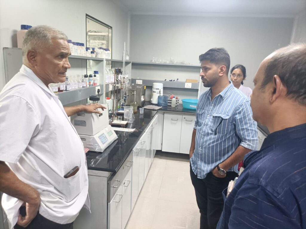
Stallen recently had the privilege of hosting two of its valued key account customers from Kerala, Mr. Ajith Paul and Mr. Praveen, for an exclusive visit to the company’s manufacturing facilities in Palghar, near Mumbai. The visit was organized with the objective of giving the customers a first-hand understanding of the company’s manufacturing excellence, quality assurance systems, and commitment to delivering world-class animal health and nutrition solutions.
The visit began at the company’s Feed Additives Manufacturing Facility located at Chahade, Palghar. During the tour, the customers were taken through various sections of the production unit, where they observed the manufacturing processes, operational systems, and quality protocols followed by the company. The technical and production teams explained the stringent standards maintained at every stage of production to ensure consistency, safety, and superior product quality.
A major highlight of the visit was the interaction at the quality control and testing laboratories, where the customers were introduced to the advanced testing procedures and monitoring systems used to maintain high manufacturing standards. The visitors showed keen interest in understanding the company’s quality assurance practices and appreciated the systematic approach followed throughout the facility.
The second part of the visit took place at Stallen’s Therapeutics Manufacturing Facility at Nadore, Palghar, one of the company’s most advanced and modern animal pharmaceutical manufacturing units. The facility, which operates under Australian GMP accreditation along with several other globally recognized quality certifications, left a strong impression on the visiting customers.
As they toured the therapeutics plant, Mr. Ajith Paul and Mr. Praveen were introduced to the sophisticated infrastructure, modern production systems, and strict compliance measures maintained for animal pharmaceutical manufacturing. The customers were also shown the advanced laboratory and testing facilities where rigorous quality checks are conducted to ensure the safety, efficacy, and reliability of every product manufactured at the facility.

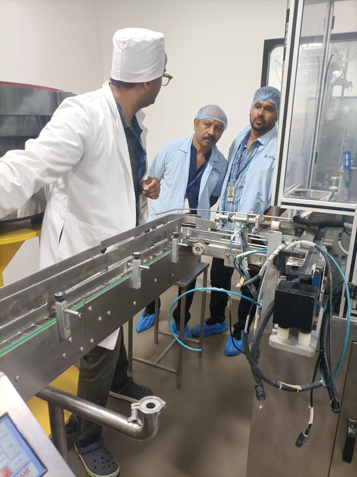

Throughout the visit, the customers appreciated the company’s transparency, professionalism, and strong focus on quality. The interaction provided them with a deeper understanding of the extensive processes, technological capabilities, and quality standards that form the foundation of Stallen’s operations.
The visit concluded on a highly positive note, with both customers expressing satisfaction and admiration for the scale, modernization, and quality-driven approach of the company’s manufacturing facilities. Such visits continue to strengthen the relationship between Stallen’s and its valued customers while reinforcing the company’s commitment to excellence in animal health and nutrition.

Date: 15.04.2026, Kochi, Kerala, India.
The Annual Conference 2025–2026 was successfully held at Vivanta by Taj, Kochi, Kerala, bringing together our sales and head office teams for three days of strategic alignment, recognition, and collaboration.
The conference commenced with an inspiring address by our Director, Mr. Aniket Parikh, who set the tone for the event with a forward-looking vision and words of motivation for the entire team. His message emphasized growth, commitment, and the opportunities that lie ahead.
A key highlight of the conference was the recognition and award ceremony, where top performers were honored with trophies and certificates. Their achievements were celebrated as a testament to dedication, excellence, and consistent performance.
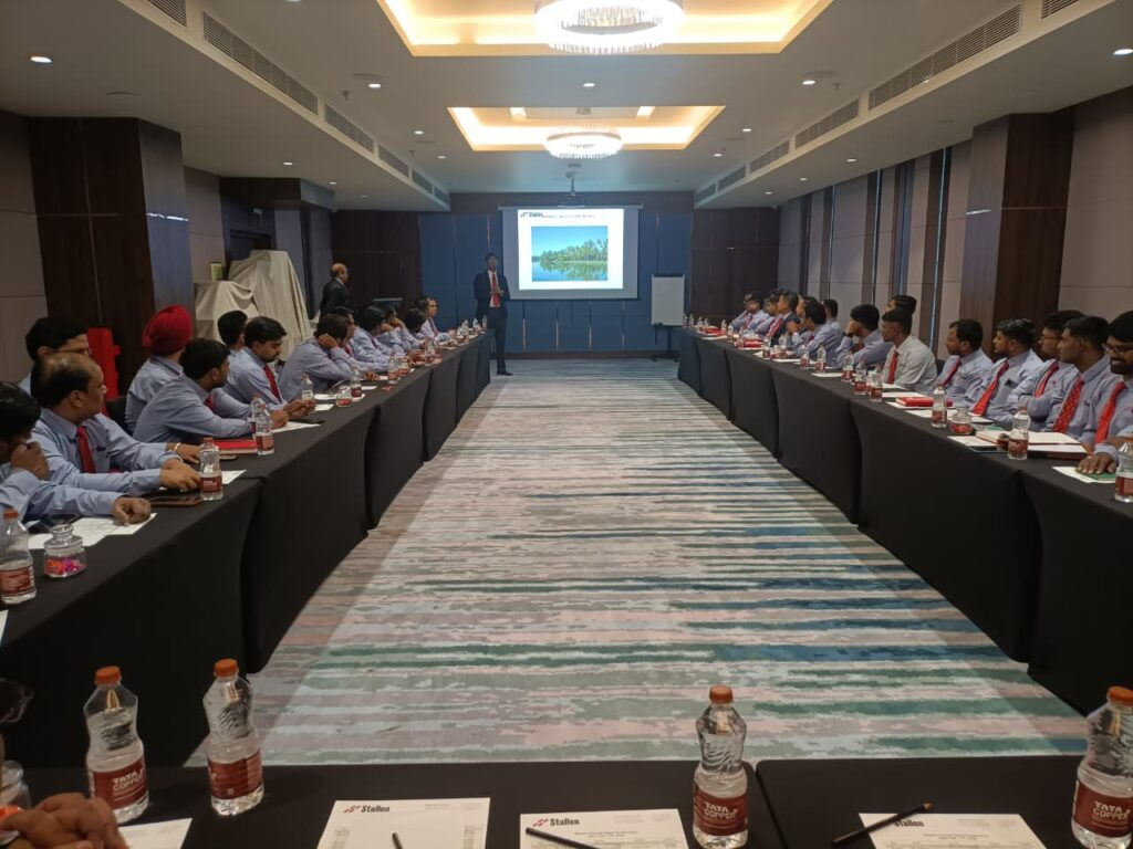

The strategic direction of the organization was further strengthened through a comprehensive session by our Chief Operating Officer, Dr. Sanjay Singhal. His presentation provided an in-depth review of the past year’s performance and outlined a clear, actionable roadmap for the upcoming year, aligning teams toward shared business objectives.
The second day was dedicated to technical excellence and innovation. Two intensive technical training sessions were conducted by our two Product Managers, along with the launch of five new products—three in the therapeutic segment and two in feed additives. These sessions were highly interactive, focusing on strong product positioning and equipping the sales team with in-depth technical knowledge for effective market execution.


To balance learning with engagement, the day also included fun and competitive team-building activities that energized participants and fostered stronger regional team bonding.
The conference concluded with one-on-one strategic discussions between the sales team and management. These interactions were instrumental in refining plans, setting targets, and aligning strategies for the upcoming year.
As the conference came to a close, it marked not just the end of the 2025–2026 cycle, but the beginning of a renewed commitment. The team departed with clear goals, strengthened strategies, and a shared promise—to return with flying colors and successfully achieve the targets and strategic objectives set for 2026–2027.


Dr. Amit V. Janbandhu & Dr. Sanjay Singhal
1. Introduction
Animal fats and vegetable oils are commonly incorporated into poultry diets due to their high energy density, which supports optimal growth performance (Blanch et al., 1996). Dietary fats and oils provide approximately 2.25 times more metabolizable energy than carbohydrates and serve as important sources of essential fatty acids and fat‑soluble vitamins. In recent years, rising feed costs have increased interest in maximizing dietary fat utilization to enhance energy concentration and meet the demands of high‑performing birds. However, fat absorption efficiency is age‑dependent; young broilers exhibit a physiological limitation in lipid digestion and absorption, which improves progressively with advancing age (Kussaibati et al., 1982)
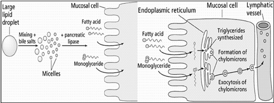
Enzymatic hydrolysis of lipids (oils and fats) produces fatty acids (FA) which are water insoluble. FA passes through the liquid phase of the small intestine and, after aggregating to form micelles, are absorbed as hydrophobic components. This process is naturally mediated by endogenous emulsifiers, such as bile salts. The assimilation of dietary fats in young birds is poor because they have a limited capacity to produce and secrete bile salts and lipase until their gastrointestinal tract matures at 10-14 days of age (Noy and Sklan,1998).
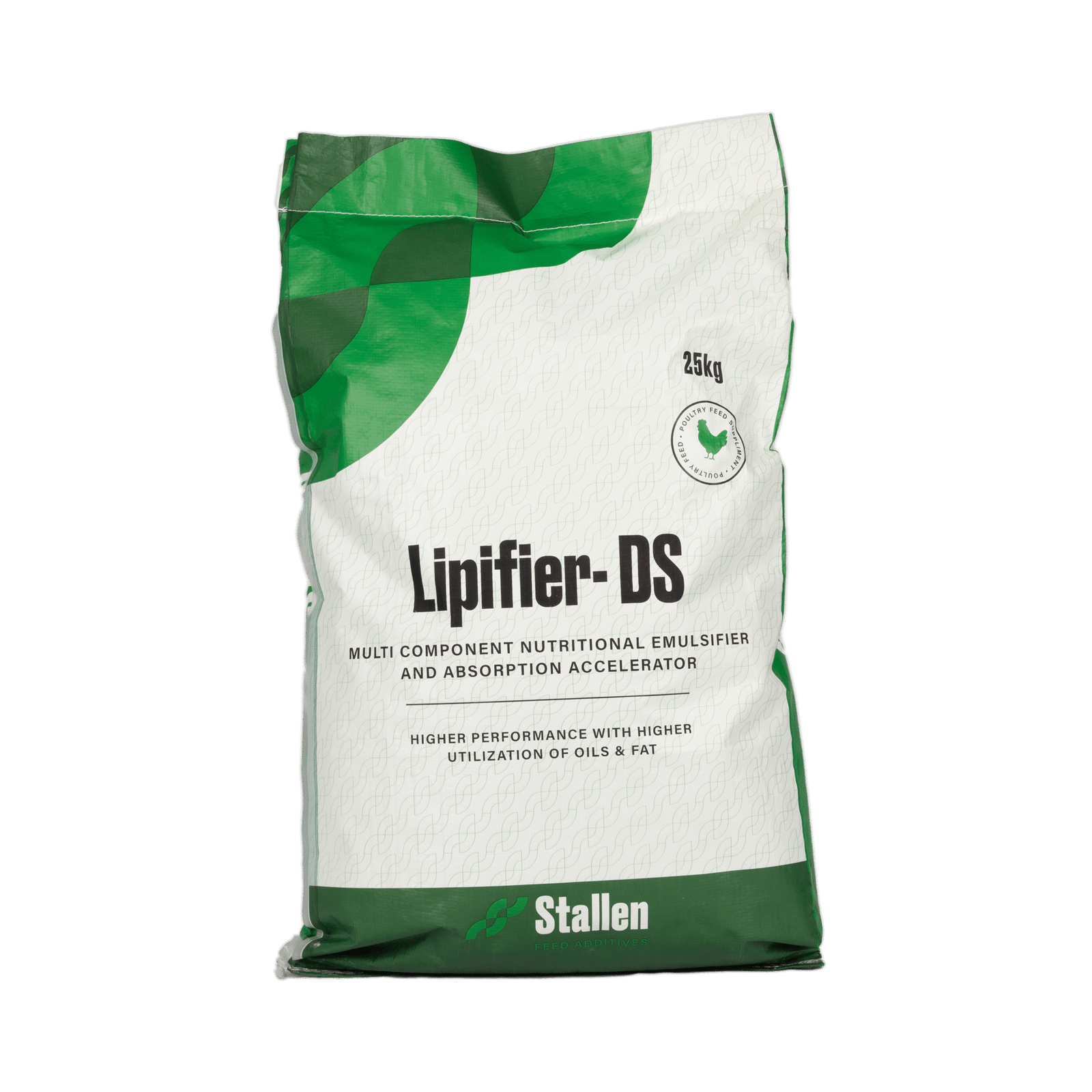
This review evaluates the role and application of exogenous emulsifiers, such as Lipifier-DS from Stallen South Asia Pvt Ltd., a multi-component emulsifier and absorption accelerator, in nutrient-dense broiler diets to maximize the growth potential of modern poultry genetics.
2. Digestibility problems in young chicks
Young birds exhibit limited fat absorption due to low endogenous lipase activity, reduced bile secretion, and poor emulsification capacity; however, these functions improve with age and adapt to higher levels of unsaturated fatty acids (Meng et al., 2004). Consequently, immature digestive systems are unable to efficiently form mixed micelles in the intestinal lumen, leading to reduced fat digestion and nutrient absorption (Leeson and Atteh, 1995). Age‑related differences in metabolisable energy (ME) utilization and growth performance have therefore stimulated interest in the use of exogenous emulsifiers to enhance fat utilisation in young birds (Roy et al., 2010). High dietary inclusion of saturated fats in broiler rations can lead to excessive visceral and carcass fat deposition, reduced vitamin A and E availability, and compromised meat quality (Chae et al., 2006).
3. Energy efficiency of emulsifiers in High‑Performance Broiler Diets
Energy is a major cost component factor in diets of high-performance animals, such as broilers. Emulsifiers can be used to improve fat digestibility and energy efficiency. As a result, lower energy diets can be formulated for birds whilst maintaining the same performance, leading to lower feed cost and more economical and sustainable production. Emulsifiers facilitate the formation of emulsion droplets, which lowers the surface tension (Ashraf, 2007), stimulates the formation of micelles, causes high levels of monoglycerides in the intestine and facilitates the nutrient transport through the membrane (Melegy et al., 2010). Emulsifier supplementation has been shown to improve feed efficiency, lipid absorption, and blood lipid profiles, although its effects on growth performance and carcass traits are inconsistent (Udomprasert and Rukkwamsuk, 2006).
4.Classification and Functional Role of Exogenous Emulsifiers in Broilers
Exogenous emulsifiers used in animal nutrition are broadly classified as natural or synthetic, with natural emulsifiers—including bile salts, phospholipids, and dietary sources such as soy lecithin—and synthetic emulsifiers comprising chemically modified molecules such as lysolecithin or lysophosphatidylcholine (Zhang et al., 2011). By modifying hydrophobic interfaces and promoting mixed‑micelle formation, these emulsifiers enhance fat digestibility, particularly in young birds with limited endogenous emulsification capacity (Al‑Marzooqi and Leeson, 1999). Soy lecithin remains the most extensively validated natural emulsifier in poultry, improving fat utilization, growth performance, and serum lipid profile while supplying choline to prevent perosis (Polin, 1980; Siyal et al., 2017; Schaible, 1970). Synthetic emulsifiers, including polyethylene glycol mono‑ and dioleates and sodium stearoyl‑2‑lactylate, have also been shown to improve growth performance and the utilization efficiency of fat, protein, and metabolizable energy, although some synthetic polyoxyethylene glycol emulsifiers exhibit lower in vivo efficiency compared with bile salts (Frobish et al., 1969; Roy et al., 2010).
5. Emulsifying agents
An emulsifying agent stabilizes an emulsion by reducing interfacial tension between immiscible phases such as oil and water, thereby preventing droplet coalescence. Emulsifiers possess both hydrophilic and lipophilic moieties, enabling their adsorption at the oil–water interface and stabilization of dispersed fat droplets. Efficient fat emulsification is a prerequisite for lipid digestion in the gastrointestinal tract and is influenced by fatty acid chain length, triglyceride structure, and degree of saturation (Gu and Li, 2003). Exogenous emulsifiers enhance lipid utilization, particularly of animal fats, and partially compensate for limited bile production and enterohepatic recirculation in young birds. Although bile salts and monoglycerides function as natural emulsifiers, their emulsification capacity is insufficient in young birds, resulting in poor fat digestibility. Furthermore, saturated and free fatty acids exhibit a lower capacity for micelle formation than long‑chain unsaturated fatty acids, further limiting lipid digestion efficiency.
Table 1. Available Emulsifiers Used in the Poultry Industry
| Category | Emulsifier | Major Effects | References |
| Natural emulsifiers | Soy lecithin | Improved growth performance; increased HDL; reduced triglycerides, insulin (INS), thyroid‑stimulating hormone (TSH), total cholesterol (TC), and LDL levels | Siyal et al., 2017; Huang et al., 2007 |
| Milk‑derived casein | Increased weight gain, improved feed conversion ratio (FCR), enhanced pancreatic lipase activity, improved ether extract digestibility, with no effect on carcass traits or serum cholesterol | Guerreiro et al., 2011 | |
| Lysophosphatidylcholine / Lysolecithin | Improved weight gain and FCR; increased digestibility of total tract apparent digestibility (CTTAD) of C16:0, C18:2, and C18:3n‑3 fatty acids; improved carcass quality and dressing percentage | Melegy et al., 2010; Zhang et al., 2011; Azman and Ciftci, 2004; Ashraf, 1995 | |
| Bile salts | Increased relative organ weights; significantly improved body weight gain (BWG); decreased plasma cholesterol; increased metabolisable energy; enhanced digestive enzyme activity in the intestinal tract | Abd‑El‑Rauof, 1995; Alzawqari et al., 2010; Gomez and Polin, 1976; Kussaibati et al., 1982; Zhong and Xiang, 2008 | |
| Synthetic emulsifiers | Glycerol polyethylene glycol ricinoleate (E 484) | Improved performance; increased intake and utilization efficiency of fat, crude protein (CP), and metabolisable energy (ME) | Roy et al., 2010 |
| Lipex® (commercial emulsifier) | Increased body weight gain and liver weight | Yordan et al., 2013 | |
| Sodium stearoyl‑2‑lactylate (SSL) | Improved body weight and relative organ weights | Flores et al., 2007; Chronakis et al., 2004 |
6. Principles of Emulsification
Emulsifiers lower the interfacial energy between the two immiscible liquids, thereby helping the formation of an emulsion. For an emulsifier to be effective at reducing droplet size and stabilising an emulsion, it needs to be located at the interface. The emulsifier must not be too soluble in either phase, otherwise it will migrate to that phase. If the emulsifier migrates away from the interface, the emulsion is destabilised.
An emulsion is most accurately defined as a dispersion of liquid droplets in a second immiscible liquid. Temporary emulsions may be formed by mixing/agitating the two normally immiscible liquids; however, the stability of temporary emulsions produced in this way is poor. Emulsifiers are surface active materials (surfactants) that are used to assist in the formation of an emulsion and to stabilise the emulsion.
Fig.1. Below is a simplistic view of an emulsion particle, protected by surfactant molecules partitioned at the interface of the internal and external phase.
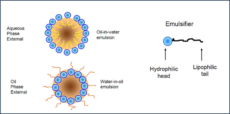
There are several different types of emulsions. They are loosely described by their phase relationship and/or by their appearance:
The appearance of the emulsion is dependent upon the particle size of the discontinuous phase.
Table.2. Particle size is listed in nanometers (nm)
| Particle size | Appearance |
| > 1 | White |
| 0.1 -1.0 | Blue White |
| 0.05 – 0.1 | Translucent |
| < 0.05 | Transparent |
7. Mechanism of Action of Lipid Digestion and Absorption
Fig.2. General schematic of lipid digestion and absorption
8. Hydrophilic-lipophilic balance
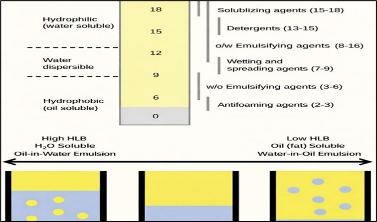
The combination of hydrophilic and lipophilic characteristics in one molecule gives it the distinctive property that the emulsifier can dissolve in fat as well as in water, and can aid in mixing the two fractions. The key indicator for selecting an emulsifier is hydrophilic-lipophilic balance (HLB), ranging from 0 to 20 which reveals the degree of fat or water solubility. Lower HLB indicates a more lipophilic or fat-soluble emulsifier. On the other hand, higher HLB indicates a more water soluble or hydrophilic emulsifier. Ideally, the emulsifier should be soluble in the continuous phase as the Bancroft rule states (1912). For the soluble condition known as the ‘fat-rich environment’ mixed in a small amount of water, an emulsifier with a lower HLB is advised, and vice versa. Because of birds consume water 1.5-2 times more than feed; the diet should contain a small amount of fat and the water amount should exceed fat in digestive tract. In this situation, a high HLB is more appropriate.
Fig. 3. HLB scale and its influence on surfactant functionality and emulsion types (Al-Yami et al. 2018)
9. Lipifier‑DS: –
Lipifier‑DS contains a blend of hydrolysed phospholipids—including LPC, LPE, LPI, LPA, other lysophospholipids—along with glyceryl polyethylene glycol ricinoleate coating.
a) Mechanism of Action of Lipifier-DS
Lipifier-DS function based on their solubility. If the emulsifier is more soluble in water, it forms an oil-in-water (O/W) emulsion, ideal for fat digestion in poultry. Conversely, if it is more soluble in oil, it forms a water-in-oil (W/O) emulsion. The emulsification process helps break down fat droplets into smaller micelle particles that remain dispersed in the water phase, allowing digestive enzymes to act more efficiently and resulting in availability of extra metabolizable energy to the birds. Exogenous emulsifiers are molecular surfactants with both hydrophobic and hydrophilic properties. The hydrophobic end with fatty acids is directed to the oil phase, while the hydrophilic end with sucrose, glycol, glycerol, sorbitol or polyglycerol is directed to the aqueous phase, forming a “molecular bridge” by decreasing surface tension that inhibits the coalescing of hydrolysed lipid droplets into large molecules.
b) Key Features of Lipifier-DS
c) Benefits of Lipifier-DS
10. Conclusion:
Stallen South Asia Pvt. Ltd.’s has Lipifier-DS is an effective exogenous emulsifier that enhances fat utilization and nutrient absorption in poultry diets. By promoting efficient emulsification through a stable PEGR coating, it improves energy availability and feed efficiency, supporting better growth and FCR in broilers, enhanced egg performance in layers, and overall higher production efficiency.
References
BLANCH, A., BARROETA, C., BAUCELLS, M.D., SERRANO, X. and PUCHAL, F. (1996), Utilisation of different fats and oils by adult chickens as a source of energy, lipid and fatty acids. Animal Feed Science and Technology 61: 335-342.
NOY, Y. and SKLAN, D. (1998) Metabolic responses to early nutrition. Journal of Applied Poultry Research 7: 437-451.
Kussaibati R, Guillaume J and Leclercq B. 1982. The effects of age, dietary fat and bile salts, and feeding rate on apparent and true metabolisable energy values in chickens. British Poultry Science 23(5): 393–403.
Meng X, Slominski B A and Guenter W. 2004. The effect of fat type, carbohydrase, and lipase addition on growth performance and nutrient utilization of young broilers fed wheat-based diets. Poultry Science 83(10): 1718–27.
LEESON, S. and ATTEH, J.O. (1995) Utilisation of fats and fatty acids by turkey poults. Poultry Science 74: 2003-2010.
ROY, A.S., HALDAR, S., MONDAL, T. and GHOSH, K. (2010) Effects of supplemental exogenous emulsifier on performance, nutrient metabolism, and serum lipid profile in broiler chickens. Veterinary Medicine International: Art. ID 262604.
ASHRAF, M. (2007) Use of Emulsifiers in High Fat Level Diets of Broilers. Doct thesis, Dept Animal Production, Faculty of Agriculture, Al Azhar University, Cairo, Egypt, 235, 2007.
MELEGY, T., KHALED, N., EL-BANA, R. and ABDELLATIF, H. (2010) Dietary fortification of a natural biosurfactant, lysolecithin in broiler. African Journal of Agriculture Research 5: 2886-2892.
UDOMPRASERT, P. and RUKKWAMSUK, T. (2006) Effect of an exogenous emulsifier on growth performance in weanling pigs. Kasetysart Journal of Natural Science 40: 652-656.
Zhang B, Haitao L, Zhao D, Guoand Y and Barri A. 2011. Effect of fat type and lysophosphatidylcholine addition to broiler diets on performance, apparent digestibility of fatty acids and apparent metabolisable energy content. Feed Science and Technology 163: 177–84.
AL-MARZOOQI, W. and LEESON, S. (1999) Evaluation of dietary supplements of lipase, detergent and crude porcine pancreas on fat utilisation by young broiler chicks. Poultry Science 78: 1561-1566.
Siyal F A, Babazadeh D, Wang C, Arain M A, Saeed M, Ayasan T, Zhang L and Wang T. 2017. Emulsifiers in poultry industry- A review. World Poultry Science Journal 73: 1–6.
Schaible P J. 1970. Poultry: Feeds and Nutrition. 2nd Edn., The AVI Publishing Co. USA.
Polin D and Hussein T H. 1982. The effect of bile acid on lipid and nitrogen retention, carcass composition, and dietary metabolizable energy in very young chicks. Poultry Science 61(8): 1697–1707.
FROBISH, L.T., HAYS, V.W., SPEER, V.C. and EWAN, R.C. (1969) Effect of diet form and emulsifying agents on fat utilisation by young pigs. Journal of Animal Science 29: 320-324.
CHAE, B.J., LOHAKARE, J.D. and CHOI, J.Y. (2006) Effects of incremental levels of α-tocopherol acetate on performance, nutrient digestibility and meat quality of commercial broilers. Asian-Australian Journal of Animal Sciences 19: 203-208.
GU, X. and LI, D. (2003) Fat nutrition and metabolism in piglets: A review. Animal Feed Science and Technology 109: 151-170.
BANCROFT, W.D. (1912) The theory of emulsification VI. The Journal of Physical Chemistry 17: 501-519.
Dr. Amit V. Janbandhu & Dr. Sanjay Singhal
1. Introduction
The poultry industry is among the most efficient sectors of agriculture, making a significant contribution to livelihood generation and global nutritional security. The global poultry population currently exceeds 26.8 billion birds (FAO, 2020). From 1961 to 2019, worldwide poultry meat production increased to approximately 132 million tonnes annually, representing nearly 37% of total global meat production (FAO, 2020). This growing demand for animal‑derived foods is primarily driven by rapid population growth, rising income levels, and increasing urbanization (FAO, 2020).
To satisfy the rising demand for meat and eggs, modern poultry production systems operate under intensive rearing conditions, exposing birds to continuous physiological stress. As a result, antibiotics have been extensively used for disease prevention, growth promotion, and immune enhancement. However, the indiscriminate use of antibiotics has hastened the emergence of antimicrobial resistance among pathogenic bacteria (Garcia‑Migura et al., 2014; Roth et al., 2019). The World Health Organization has identified antimicrobial resistance as “a serious threat to public health worldwide that requires action across all government sectors and society” (WHO Factsheets, 2015). Antibiotic‑resistant bacteria can enter the human food chain through animal‑derived products, posing substantial public health risks (Cui et al., 2005). Moreover, fresh meat products may act as reservoirs of antibiotic‑resistance genes that can be transferred to humans through regular consumption (Diarrassouba et al., 2007). In response to the escalating challenge of antimicrobial resistance, the livestock industry is increasingly exploring effective alternatives to conventional antibiotics, with probiotics emerging as a promising and sustainable solution.
2. What Is Probiotics?
The concept of probiotics was first introduced in the early 1900s by the Russian‑born Nobel laureate Elie Metchnikoff, who demonstrated that the regular consumption of beneficial microorganisms could positively influence gastrointestinal health (Metchnikoff, 1907). His observations of populations that consumed fermented milk products led to the hypothesis that beneficial intestinal microflora enhance resistance to pathogenic organisms. The term probiotics is derived from Greek, meaning “pro‑life” (Shokryazdan et al., 2017a, 2017b). According to the FAO/WHO, probiotics are defined as “live organisms which, when administered in adequate amounts, confer a health benefit on the host” (FAO/WHO Joint Report, 2001).
Probiotics are widely recognized for their ability to modulate gut microflora and enhance immune responses (Chen et al., 2012) and are extensively applied in both clinical and veterinary practices (Abushelaibi et al.). In livestock production, probiotic supplementation has been associated with improved growth performance, production efficiency, disease resistance, nutrient digestibility, immune function, and fecal microbial balance (Lan et al., 2017). In recent years, non‑specific immunomodulators—including probiotics, prebiotics, synbiotics, postbiotics, polysaccharides, organic acids, enzymes, and essential oils—have gained considerable attention as alternatives to conventional antibiotics and are increasingly used to promote gut health in poultry birds (Callaway et al., 2017).
3. Probiotic and Related Biotic Agents in Poultry
The species currently being used in probiotic preparations are varied and many. These are mostly Lactobacillus bulgaricus, Lactobacillus acidophilus, Lactobacillus casei, Lactobacillus helveticus, Lactobacillus lactis, Lactobacillus salivarius, Lactobacillus plantarum, Streptococcus thermophilus, Enterococcus faecium, Enterococcus faecalis, Bifidobacterium spp. and Escherichia coli. With two exceptions, these are all intestinal strains. The two exceptions, Lactobacillus bulgaricus and Streptococcus thermophilus, are yoghurt starter organisms (Fuller et.al, 1989). Some other probiotics are microscopic fungi such as strains of yeasts belonging to Saccharomyces cerevisiae species (Guillot et.al, 1998).
In broiler nutrition, probiotic species belonging to Lactobacillus, Streptococcus, Bacillus,
Bifidobacterium, Enterococcus, Aspergillus, Candida, and Saccharomyces have a beneficial effect on broiler performance (Tortuero et.al, 1973), modulation of intestinal microflora and pathogen inhibition, intestinal histological changes, immunomodulation, certain haematobiochemical parameters, improving sensory characteristics of dressed broiler meat and promoting microbiological meat quality of broilers (Kabir et.al, 2005).
The International Scientific Association of Probiotics and Prebiotics defines prebiotics as “a substrate that is selectively utilized by host microorganism conferring a health benefit” (Gibson et al., 2017). Synbiotics are defined as a “synergistic combination of probiotics and prebiotics that are beneficial for the host by improving the development and colonization of live microorganisms in the gut” (FAO/WHO joint report, 2002). Postbiotics are defined as “the preparation of inanimate microorganism and/or their components that confer a health benefit on the host” (Salminen et al., 2021).
Given their safety, efficacy, and sustainability, probiotics have gained considerable attention as viable alternatives to antibiotics. This article outlines the status, mechanisms, and use of probiotics in poultry as alternatives to antibiotics.”
4. The concept of probiotics
In healthy, non-stressed poultry, a dynamic balance exists between beneficial and non-beneficial gut bacteria, which is essential for optimal performance. Stress disrupts this balance by reducing beneficial flora, particularly lactobacilli, allowing overgrowth of harmful microorganisms. This imbalance may result in clinical conditions such as diarrhea or subclinical effects that impair growth and feed efficiency. Although the protective gut microflora is relatively stable, it can be influenced by key factors including excessive hygiene, antibiotic use, and stress. Under natural conditions, chicks acquire a complete and protective gut microflora through contact with the hen, providing resistance against infection. In contrast, commercially reared chicks hatch in sanitized incubators lacking normal intestinal microorganisms. Gut colonization may be influenced by eggshell microbiota and the onset of gastric HCl secretion at approximately 18 days of incubation, which affects microbial selection. Consequently, early probiotic supplementation is particularly important in poultry, as chicks are deprived of maternal microbial transfer and can benefit from microbial preparations that restore protective gut microflora (Fuller et al., 2001).
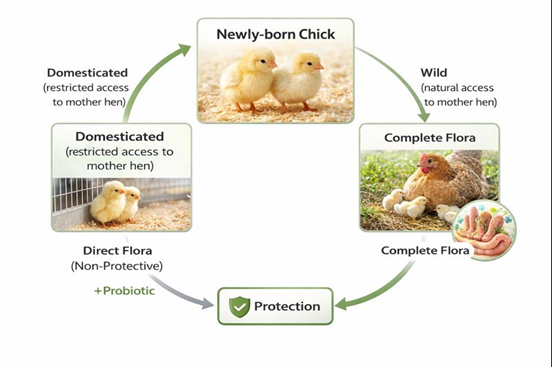
Figure 1. Schematic representation of the concept of probiotics (modified from (Fuller et.al, 2001).
5.Mechanism of action of probiotics
Probiotics place a key role in gut microbial health. The mechanisms of action of probiotics mainly include two, i.e. competitive exclusion and immune system modulation.
Competitive exclusion of pathogens by probiotics includes:
(a) production of inhibitory compounds like bacteriocins, mucins, defensins, etc. (b) preventing the adhesion of pathogens, (c) competition for nutrients, (d) reduction of toxin bioavailability, and (e) modulation of the host immune system including the enhancement of both innate and adaptive immunity (Hernandez-Patlan et al., 2020).
a) Secretion of inhibitory compounds
Probiotics inhibit pathogenic bacteria through the production of antimicrobial compounds, including antimicrobial peptides (AMPs) such as bacteriocins, as well as organic acids, hydrogen peroxide, ethanol, diacetyl, and carbon dioxide (Liao & Nyachoti, 2017). Bacteriocins are ribosomally synthesized AMPs that suppress pathogens by disrupting cell wall synthesis or forming membrane pores while sparing beneficial gut microbiota (Cotter et al., 2013). Pediocin A from Pediococcus pentosaceus, divercin from Carnobacterium divergens, and nisin from Lactococcus lactis have shown inhibitory effects against Clostridium perfringens and improved broiler performance (Grilli et al., 2009). Synergistic bacteriocin activity with other biomolecules has been reported against multiple pathogens (Rishi et al., 2014). Organic acids reduce intracellular pH and disrupt bacterial metabolism and membranes, while lactic acid bacteria inhibit Salmonella, Listeria monocytogenes, and Escherichia coli without harming intestinal epithelium (Ricke, 2003). Additional inhibitory effects are mediated by ethanol, diacetyl, and carbon dioxide (Ingram, 1989).
b) Inhibition of pathogenic adhesion
Probiotics prevent pathogen colonization by competitively blocking adhesion sites on intestinal epithelial cells, a key criterion for selecting effective probiotic strains . Probiotic adhesion stimulates mucosal immunity and promotes the secretion of mucins and defensins, thereby strengthening the epithelial barrier (Bermudez-Brito et al., 2012). Mucins are highly glycosylated glycoproteins that form the mucus layer and inhibit pathogen attachment and colonization (Collado et al., 2005). Interactions between probiotic surface proteins and intestinal epithelial cells further exclude pathogens. Defensins, small cationic antimicrobial peptides, inhibit bacterial growth by disrupting membranes or cell wall synthesis and can neutralize bacterial toxins (Ayabe et al., 2000).
c) Competition for nutrients
Probiotics limit pathogen growth by competing for essential nutrients and occupying intestinal epithelial adhesion sites, thereby restricting pathogen attachment in the gastrointestinal tract . This competitive exclusion reduces pathogen proliferation and colonization and creates unfavorable conditions for pathogen survival (Callaway et al., 2008). Competitive exclusion has been demonstrated in vitro using chicken intestinal mucosa (Hirn et al., 1992). In vivo studies show that early supplementation with lactobacillus-based probiotics (1 × 10⁵ CFU/mL, 1–7 days of age) significantly reduced Salmonella colonization in chicks (Penha Filho et al., 2015).
d) Reduction in toxin bioavailability
Probiotics like lactobacillus help the reduction in the uptake of pathogenic toxins in the intestinal cells. The positive effects of LAB-based probiotics had helped in the reduction of toxin expression in the gut. Lactic acid bacteria are known for their natural barriers against mycotoxins which are harmful compounds for animals. A few strains can also eradicate the detrimental reactions of aflatoxins on human and animal health (Abbes et al., 2016).
e) Modulation of the host immune system
Probiotics modulate host immunity by interacting with intestinal epithelial cells, dendritic cells, macrophages, and lymphocytes. These interactions enhance innate immune defenses by limiting pathogen proliferation and reinforcing epithelial barriers through increased mucus and antimicrobial peptide production. Intestinal epithelial and dendritic cells recognize probiotics via pattern recognition receptors, initiating immune signaling cascades. Activation of antigen-presenting cells stimulates adaptive immunity through T- and B-cell responses. Probiotics regulate cytokine expression and suppress intestinal inflammation by downregulating TLR and NF-κB signaling pathways. Enhanced IgA and IgG responses have been observed in broilers supplemented with probiotic strains such as Clostridium butyricum and Lactobacillus plantarum (Han et al., 2018).
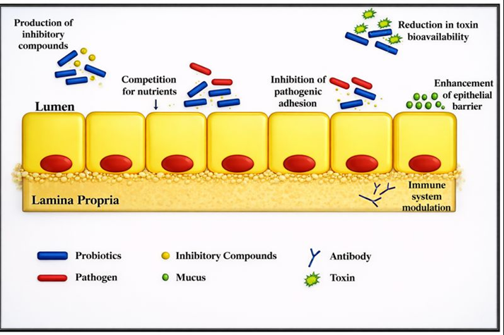
Fig. 2. Mode of action of probiotics. It starts with the secretion of inhibitory compounds leading to inhibition of the pathogen adhesion to the epithelial layer of the GI tract besides creating competition for nutrients among pathogens thereby reducing their colonization. Also, it helps in diminishing the toxin bioavailability and modulates the immune system of the host by activating adaptive and innate immunity.
6. Single- and multi-strain probiotics
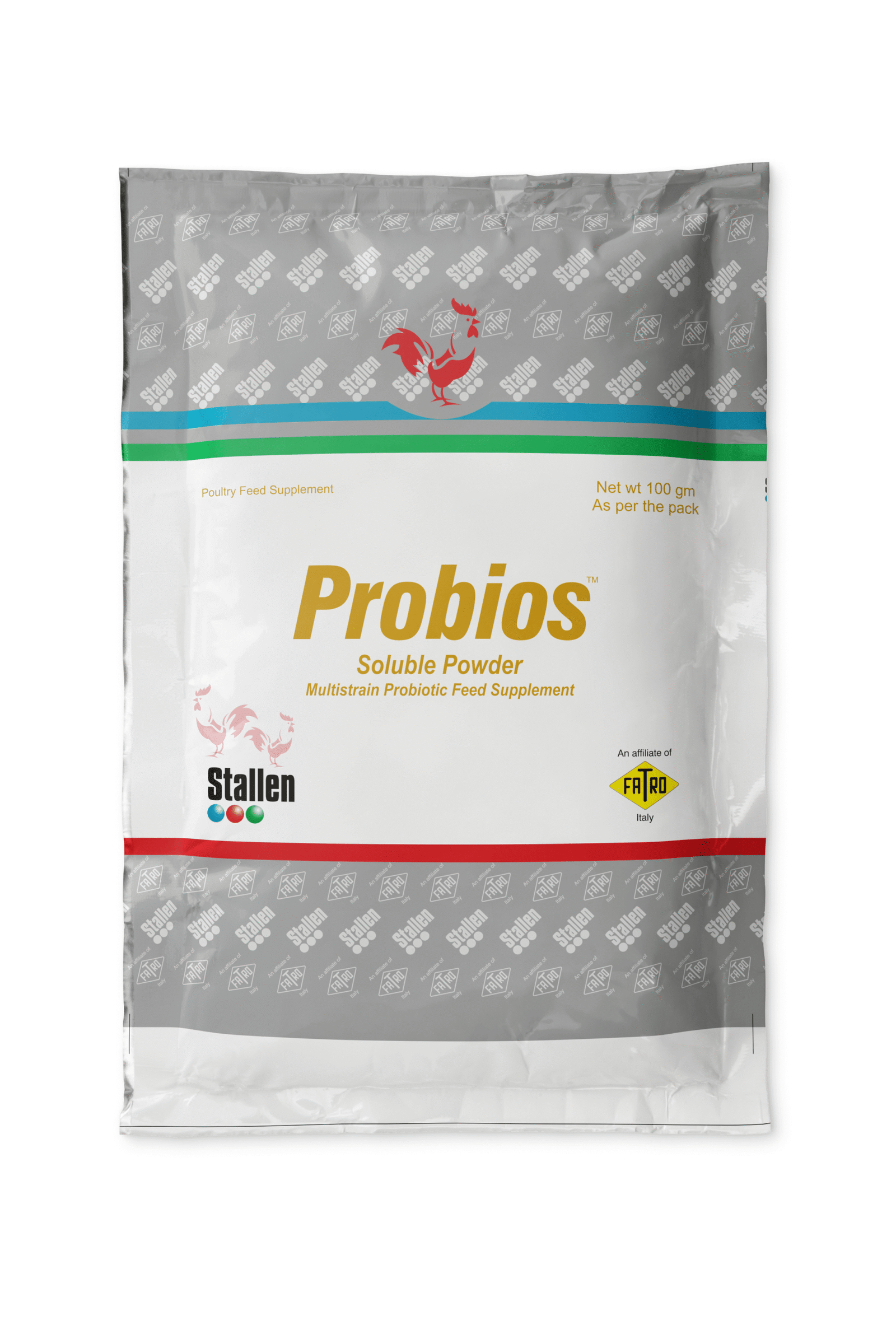
Probiotics are classified as single- or multi-strain formulations. Single-strain probiotics contain one microbial species, commonly Lactobacillus, Bifidobacterium, Streptococcus, Pediococcus, Enterococcus, Bacillus, Saccharomyces, and Micrococcus. Multi-strain probiotics combine multiple strains or genera to provide complementary benefits and have been shown to improve growth performance and gut health in broilers, including under disease challenge conditions. Commercial products such as Probios from Stallen South Asia private Ltd contain diverse probiotic combinations. Probiotic efficacy depends on strain composition and viable counts, with variable outcomes reported (Aalaei et al., 2018).
a) Bacillus
Many strains of Bacillus have potential against pathogenic bacteria. A group of researchers isolated 200 Bacillus strains from the faeces of broiler chicken and many strains among them showed activity against C. perfringens in in vitro conditions. A study suggested that B. subtilis strain SP6 when used in a field trial, the mortality of chicken infected with Necrotic enteritis was reduced to half. It also reduced the number of C. perfringens and enhanced the intestinal health of chickens. Regular use of B. licheniformis supplementation reduced mortality and increased the performance among the chicks (Knap et al., 2010).
b) Yeast
Yeasts possess antimicrobial and immunomodulatory properties, largely due to β-glucans that stimulate host immunity. They inhibit pathogens by producing mycocins, degrading toxins, preventing epithelial adhesion, and competing for nutrients. Saccharomyces boulardii improves intestinal health and reduces Salmonella enteritidis infection, while recombinant Pichia pastoris expressing Clostridium perfringens α-toxin enhances broiler performance (Gil de Lossantos et al., 2005).
c) Enterococci
Enterococci produce bacteriocins (enterocins) with activity against Gram-positive and Gram-negative bacteria . Enterococcus faecium supplementation reduces Clostridium perfringens, alleviates coccidiosis, and improves growth performance and nutrient utilization in broilers. Enhanced IgA production, immune responses, and microbiome modulation have also been reported with E. faecium and E. faecalis supplementation (Beirão et al., 2018)
7. Beneficial effects of probiotics on poultry
a) Effects on growth performance and productivity
Probiotics improve body weight gain, feed intake, feed conversion ratio, and overall productivity in poultry. Supplementation with Pediococcus acidilactici and Bacillus subtilis enhances egg quality, increases eggshell thickness, and reduces yolk cholesterol. Multi-strain probiotics improve egg production and mitigate heat-stress effects. Probiotics also enhance meat microbiological quality, reduce Salmonella enteritidis contamination, and improve nutrient metabolism and growth performance (Bailey et al., 2000).
b) Effects on serum biochemistry
Probiotic supplementation significantly modulates serum biochemistry in poultry by reducing total cholesterol, LDL, VLDL, triglycerides, uric acid, and liver enzymes (ALT, AST), while increasing protein and calcium levels . Lactobacillus spp., Enterococcus faecium, and Bacillus subtilis reduce cholesterol absorption and improve lipid metabolism in broilers and layers (Kalavathy et al., 2003).
c) Effect on health and immunity
Probiotics enhance poultry health and immunity by modulating gut microbiota and immune signaling. Lactic acid bacteria (LAB) regulate pro- and anti-inflammatory cytokines (IL-1β, IL-6, IL-10, IFN-γ, TNF-α) and suppress inflammation through NF-κB and TLR-mediated pathways. Supplementation with Clostridium butyricum, Lactobacillus spp., and Saccharomyces cerevisiae enhances gut flora, T-cell responses, intraepithelial lymphocyte activity, and mucosal immunity in broilers (Yang et al., 2012).
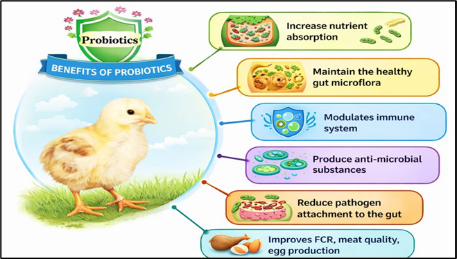
Fig. 3. Positive effects of probiotics on poultry
Table 1. Few studies showing the potentials of probiotics in poultry.
| S. No. | Probiotic species / formulation | Key findings | Reference |
| 1 | Lactobacillus spp. | Reduced Clostridium perfringens load in chicken gut | Gerard et al., 2008 |
| 2 | Lactobacillus spp. | Reduced E. coli and improved gut health | Grilli et al., 2009 |
| 3 | Lactobacillus spp. | Improved growth performance and intestinal morphology | De Cesare et al., 2020 |
| 4 | Pediococcus acidilactici | Improved egg quality and reduced yolk cholesterol | Mikulski et al., 2012 |
| 5 | Bacillus subtilis | Improved egg quality and reduced heat-stress effects | Sobczak & Kozłowski, 2015; Abdelqader, 2020 |
| 6 | Lactobacillus salivarius | Improved BWG and FCR in broilers | Shokryazdan et al., 2017a, 2017b |
| 7 | Lactobacillus plantarum | Improved gut integrity and immune response | Wang et al., 2018 |
| 8 | L. plantarum + P. polymyxa | Reduced ALT, AST, LDL, and uric acid | Wu et al., 2019 |
| 9 | Lactobacillus spp. | Reduced serum cholesterol and triglycerides | Fathi, 2013 |
| 10 | L. sporogenes | Improved serum protein and reduced lipid profile | Arun et al., 2007 |
| 11 | Lactobacillus acidophilus | Improved gut health and metabolic functions | De Cesare et al., 2020 |
| 12 | Enterococcus faecium | Reduced cholesterol and improved immunity | Capcarova et al., 2010 |
| 13 | Lactobacillus salivarius | Reduced cholesterol and LDL | Shokryazdan et al., 2017a, 2017b |
| 14 | Multi-strain probiotics | Improved gut health and performance | Taherpour et al., 2009 |
| 15 | Lactobacillus spp. | Improved nutrient metabolism and absorption | Burgain et al., 2014 |
| 16 | Lactobacillus spp. | Reduced inflammatory cytokines | Chen et al., 2012; Park et al., 2014 |
| 17 | Clostridium butyricum | Enhanced gut flora and immune response | Yang et al., 2012 |
| 18 | Lactobacillus fermentum | Improved T-cell response | Bai et al., 2013 |
| 19 | L. plantarum + L. johnsonii | Suppressed NF-κB activation | Joo et al., 2011; Li et al., 2015 |
| 20 | Lactobacillus spp. | Reduced IL-1β and enhanced TLR4 expression | Li et al., 2015 |
| 21 | Lactococcus lactis | Induced pro-inflammatory immune signaling | Slawinska et al., 2021 |
| 22 | Lactobacillus casei | Increased IELs and mucosal immunity | Tian et al., 2021 |
| 23 | Multi-strain probiotics | Improved growth and gut microbiota | Lambo et al., 2021 |
| 24 | Bacillus subtilis | Reduced C. perfringens and necrotic enteritis | Jayaraman et al., 2013 |
| 25 | Bifidobacterium spp. | Enhanced immune response | Knapp et al., 2010 |
| 26 | Yeast (Saccharomyces cerevisiae) | Improved gut health and immunity | Novak & Vetvicka, 2008 |
| 27 | Saccharomyces boulardii | Improved intestinal health | Rajput et al., 2013 |
| 28 | Enterococcus faecium | Reduced C. perfringens and improved immunity | Cao et al., 2013 |
| 29 | Enterococcus faecium | In vitro anti-C. perfringens activity | Shin et al., 2008 |
| 30 | Enterococcus faecium | Reduced coccidiosis severity | El-Sawah et al., 2020 |
| 31 | Enterococcus faecium | Improved nutrient utilization and metabolism | Zheng et al., 2016 |
Table 2. Impact of probiotics on chicken production
| Probiotic strain | Host age (condition) | Chicken type | Dosage | Duration | Results | Reference |
| Lactobacillus rhamnosus | 70 weeks | Layers | 1 × 10⁹ CFU | 88 weeks | ↑ Egg production | Huang et al., 2023 |
| Bacillus subtilis | Day-old chicks | Broiler | 500 mg/kg | 42 weeks | ↓ mortality, ↑ ADFI and BW, → FCR and ADG | Qiu et al., 2021 |
| Bacillus amyloliquefaciens | Day-old chicks | Broiler | 10⁶ CFU/g | 42 days | ↑ Body weight and average daily weight gain | Mazanko et al., 2022 |
| Pediococcus acidilactici + Lactobacillus plantarum | Day-old chicks | Broiler | 2 × 10⁹ CFU/chick | 42 days | ↑ Feed conversion and production efficiency, ↓ myopathies | Paz et al., 2019 |
| Lactobacillus acidophilus, L. plantarum + Bifidobacterium spp. | Day-old chicks | Broiler | 1.2 × 10⁹ CFU unit/mL | 21 days | ↑ BW, FCR, feed consumption (starter), ↑ breast muscles, liver, heart, kidneys, lungs | Agustono et al., 2022 |
| Lactobacillus casei, L. acidophilus + Bifidobacterium | Day-old chicks | Broiler | 10 mL probiotics/L water (oral) | 42 days | ↑ Eviscerated yield and breast yield, ↓ ADFI, FCR, abdominal fat | Zhang et al., 2021 |
| Bacillus subtilis | Day-old chicks | Broiler | 0.1% probiotics | 42 days | ↑ BW and ADWG | Popov et al., 2024 |
| Bacillus subtilis | Day-old chicks | Broiler | 1.0 × 10⁶ spores/g feed | 42 days | ↑ Bone mineralization, density, size, wall thickness, and tibia/femur weight | Yan et al., 2018 |
| Bifidobacterium spp. (in ovo) | Day-old chicks | Broiler | 2 × 10⁸ CFU | 28 days | ↑ LBW and DBWG | Abdel-Moneim et al., 2020 |
| Lactobacillus fermentum + Saccharomyces cerevisiae | Day-old chicks | Broiler | 1 × 10⁷ CFU/g and 2 × 10⁶ CFU/g | 42 days | ↑ ADG and feed efficiency | Bai et al., 2013 |
| Lactobacillus johnsonii | Day-old chicks | Broiler | 1 × 10⁵ CFU/g BS15/g | 42 days | No changes | Wang et al., 2018a |
| Lactobacillus johnsonii | Day-old chicks | Broiler | 1 × 10⁶ CFU/g BS15 | 28 days | ↑ Starter, finisher, and overall daily weight gain | Wang et al., 2018a |
| Bacillus subtilis | 25 weeks | Layers | 9.0 × 10⁵ CFU/g | 122 days | ↑ Hatchability, fertility, egg weight, yolk colour/index, eggshell thickness | Liu et al., 2019 |
| Bacillus subtilis | 28 weeks | Layers | 1.0 × 10⁵, 1.0 × 10⁶, 1.0 × 10⁷, 1.0 × 10⁸ (B4) CFU/g | 24 weeks | No significant effect on egg production | Guo et al., 2017 |
| Bacillus subtilis | 32 weeks | Layers | 4 × 10⁹ CFU/g | 90 days | No significant effect on ABWG and FCR | Fathi et al., 2018 |
8.Probios
Probios contains nine different species of beneficial microflora, each at a concentration of 2 × 10⁸ CFU. These include Bifidobacterium bifidum, Lactobacillus acidophilus, Lactobacillus bulgaricus, Lactobacillus casei, Lactobacillus plantarum, Streptococcus faecium, and Streptococcus thermophilus, along with beneficial yeasts such as Torulopsis spp. and Aspergillus spp.
a) Mechanism of action Probios :
Production of lactic acid in the gut which reduce the pH and provides unfavorable conditions for pathogenic bacteria. Probios help in production of antibacterial compounds like lysozyme, lactoferrin, lactoperoxidase and bacteriocins. These compounds are bacteriostatic and bacteriocidal in nature. It reduces toxin production by suppressing the growth and colonization of E. coli. It increases Immunostimulation by increasing macrophage and lymphocyte activity. It rigorously competes with intestinal harmful microbes by competitive exclusion mechanism which help to restrict detrimental colonization to bind with receptors in the mucus layer. It helps in barrier function by improving mucin glycoproteins secretion through mucus producing cells to yield a dense mucus layer that helps to decrease intracellular permeability to pathogens.
b) Charecteristic Of Probios
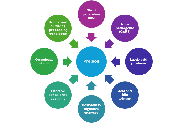
c) Benefits of Probios
d) Comparision between Probios and common probiotic
| Probios | Common Probiotic |
| Made from direct fed microbial (DFM) | Made from spores |
| Contains 9 strains of microflora i.e. multistrain | Contains 2 to 3 species of microflora |
| Curdling of milk is observed when a teaspoon of Probios is added to milk, kept overnight | No curdling of milk is observed |
| High concentration of viable cells | Low concentration of viable cells |
| More viable in GI tract | Less viable in GI tract |
| Withstands pelletization temperature | Does not withstand pelletization temperature |
| Longer shelf life | Shorter shelf life |
9.Conclusion:
Probios, has demonstrated significant positive effects on poultry health and productivity. Its supplementation enhances gut enzyme activity, protein and fat metabolism, feed efficiency, fiber digestion, and organic phosphorus utilization, leading to improved body weight gain and feed conversion ratio in broilers. Probios also supports better litter conditions, reduces stress and mortality, promotes the development of intestinal mucous glands and villi, and maintains a healthy gastrointestinal tract. In layers and breeders, it improves egg production, egg quality, and shell strength. Additionally, Probios contributes to mycotoxin detoxification without causing adverse effects, making it a safe and effective probiotic solution for sustainable poultry production.
References
FAO, 2020, Gateway to poultry production and products. http:// www. fao. org/ poult ry- produ ction- produ cts/ produ ction/ poult ry- speci es/ en/. Accessed on June 11, 2020.
Garcia-Migura, L., Hendriksen, R. S., Fraile, L., & Aarestrup, F. M. (2014). Antimicrobial resistance of zoonotic and commensal bacteria in Europe: The missing link between consumption and resistance in veterinary medicine. Veterinary Microbiology, 170, 1–9.
Roth, N., Kasbohrer, A., Mayrhofer, S., Zitz, U., Hofacre, C., & Domig, K. J. (2019). The application of antibiotics in broiler production and the resulting antibiotic resistance in Escherichia coli: A global overview. Poultry Science, 98(4), 1791–1804.
WHO Factsheets. Antimicrobial resistance. April 2015. Available at: http:// www. who. int/ media centre/ facts heets/ fs194/ en. Accessed September 14, 2015.
Cui, S., Ge, B., Zheng, J., & Meng, J. (2005). Prevalence and antimicrobial resistance of Campylobacter spp. and Salmonella serovars in organic chickens from Maryland retail stores. Applied and Environmental Microbiology, 71, 4108–4111.
Diarrassouba, F., Diarra, M. S., Bach, S., Delaquis, P., Pritchard, J., Topp, E., & Skura, B. J. (2007). Antibiotic resistance and virulence genes in commensal Escherichia coli and Salmonella isolates from commercial broiler chicken farms. Journal of Food Protection, 70, 1316–1327.
Metchnikoff, E. (1907). Lactic acid as inhibiting intestinal putrefaction. In M. P. Chalmers (Ed.), The Prolongation of Life: Optimistic Studies (pp. 161–183).
Shokryazdan, P., FaselehJahromi, M., Liang, J. B., & Ho, Y. W. (2017a). Probiotics: From isolation to application. Journal of the American College of Nutrition, 36(8), 666–676. https:// doi. org/ 10. 1080/ 07315 724. 2017. 13375 29.
Shokryazdan, P., FaselehJahromi, M., Liang, J. B., Ramasamy, K., Sieo, C. C., & Ho, Y. W. (2017b). Effects of a Lactobacillus salivarius mixture on performance, intestinal health, and serum lipids of broiler chickens. PLoS ONE, 12(5), e0175959.
Food and Agriculture Organization/World Health Organization: “Report of a Joint FAO/WHO Expert Consultation on Evaluation of Health and Nutritional Properties of Probiotics in Food Including Powder Milk with Live Lactic Acid Bacteria.” Cordoba, Argentina: Author, 2001.
Chen, C. Y., Tsen, H. Y., Lin, C., Yu, B., & Chen, C. (2012). Oral administration of a combination of select lactic acid bacteria strains to reduce the Salmonella invasion and inflammation of broiler chicks. Poultry Science,91(9), 2139–2147.
Abushelaibi, A., Al-mahadin, S., El-tarabily, K., Shah, N. P., & Ayyash, M. (2017). Characterization of potential probiotic lactic acid bacteria isolatedfrom camel milk. LWT Food Science and Technology, 79, 316–325.
Lan, R., Tran, H., & Kim, I. (2017). Effects of probiotic supplementation in different nutrient density diets on growth performance, nutrient digestibility, blood profiles, faecal microflora and noxious gas emission in weaning pig. Journal Ofthe Science of Food and Agriculture, 97, 1335–1341.
Callaway, T. R, Edrington, T. S, Byrd, J., & Nisbet, D. J. (2017). Use of direct-fed microbials in layer hen production-performance response and Salmonella control, producing safe eggs: the microbial ecology of Salmonella. Pages 301−322 in Producing Safe Eggs. S. C. Ricke and R. K. Gast, eds. Academic Press, Cambridge, MA.
Gibson, G. R., Hutkins, R., Sanders, M. E., Prescott, S. L., Reimer, R. A., Salminen, S. J., Scott, K., Stanton, C., Swanson, K. S., Cani, P. D., Verbeke, K., & Reid, G.(2017). Expert consensus document: The international scientific association for probiotics and prebiotics (ISAPP) consensus statement on the definition and scope of prebiotics. Nature Reviews Gastroenterology and Hepatology, 14, 491.
Food and Agriculture Organization of the United Nations/World Health Organization (FAO/WHO). Guidelines for the Evaluation of Probiotics in Food. In Joint FAO/WHO Working Group on Drafting Guidelines for the Evaluation of Probiotics in Food; WHO: London, ON, Canada, 2002.
Salminen, S., Collado, M. C., Endo, A., Hill, C., Lebeer, S., Quigley, E. M. M., Sanders, M. E., Shamir, R., Swann, J. R., Szajewska, H., & Vinderola, G. (2021). The international scientific association of probiotics and prebiotics (ISAPP) consensus statement on the definition and scope of postbiotics. Nature Reviews. Gastroenterology and Hepatology, 18, 649–667.
https:// doi. org/ 10. 1038/ s41575- 021- 00440-6.
Tortuero, F. Influence of the implantation of Lactobacillus acidophilus in chicks on the growth,
feed conversion, malabsorption of fats syndrome and intestinal flora. Poult. Sci. 1973, 52,
197-203.
Kabir, S.M.L.; Rahman, M.M.; Rahman, M.B. Potentiation of probiotics in promoting
microbiological meat quality of broilers. J. Bangladesh Soc. Agric. Sci. Technol. 2005, 2, 93-96.
Fuller, R. Probiotics in man and animals. J. Appl. Bacteriol. 1989, 66, 365-378.
Guillot, J.F. Les probiotiques en alimentation animale. Cah. Agric. 1998, 7, 49-54.
Fuller, R. The chicken gut microflora and probiotic supplements. J. Poult. Sci. 2001, 38, 189-196.
Liao, S. F., & Nyachoti, C. M. (2017). Using probiotics to improve swine gut health and nutrient utilization. Animal Nutrition, 3, 331–343. https:// doi. org/ 10. 1016/j. aninu. 2017. 06. 007.
Cotter, P. D., Ross, R. P., & Hill, C. (2013). Bacteriocins— a viable alternative to antibiotics? Natures Reviews Microbiology, 11, 95–105.
Grilli, E., Messina, M. R., Catelli, E., Morlacchini, M., & Piva, A. (2009). Pediocin A improves growth performance of broilers challenged with Clostridium perfringens. Poultry Science, 88, 2152–2158.
Rishi, P., Preet Singh, A., Garg, N., & Rishi, M. (2014). Evaluation of nisin-β-lactam antibiotics against clinical strains of Salmonella enterica serovar Typhi. Journal of Antibiotics, 67, 807–811.
Ricke, S. C. (2003). Perspectives on the use of organic acids and short chain fatty acids as antimicrobials. Poultry Science, 82, 632–639.
Ingram, L. O. (1989). Ethanol tolerance in bacteria. Critical Reviews in Biotechnology, 9, 305–319.
Bermudez-Brito, M., Plaza-Diaz, J., Munoz-Quezada, S., Gomez-lorente, C., & Gil, A. (2012). Probiotic mechanisms of action. Annals of Nutrition and Metabolism, 61, 160–174.
Collado, M. C., Gueimonde, M., Hernandez, M., Sanz, Y., & Salminen, S. (2005). Adhesion of selected Bifidobacterium strains to human intestinal mucus and the role of adhesion in entero pathogen exclusion. Journal of Food Protection, 68, 2672–2678.
Ayabe, T., Satchell, D. P., Wilson, C. L., Parks, W. C., Selsted, M. E., & Ouellette, A. J. (2000). Secretion of microbicidal alpha-defensins by intestinal Paneth cells in response to bacteria. Nature Immunology, 1, 113–118.
Callaway, T. R., Edrington, T. S., Anderson, R. C., Harvey, R. B., Genovese, K. J., Kennedy, C. N., Venn, D. W., & Nisbet, D. J. (2008). Probiotics, prebiotics, and competitive exclusion for prophylaxis against bacterial disease. Animal Health Research Reviews, 9(2), 217–225.
Hirn, J., Nurmi, E., Johansson, T., & Nuotio, L. (1992). Long-term experience with competitive exclusion and salmonellas in Finland. International Journal of Food Microbiology, 15, 281–285.
Penha Filho, R. A. C., Diaz, S. J. A., Fernando, F. S., Chang, Y. F., Andreatti Filho, R. L., & Junior, A. B. (2015). Immunomodulatory activity and control of Salmonella Enteritidis colonization in the intestinal tract of chickens by Lactobacillus based probiotic. Veterinary Immunology and Immunopathology, 167, 64–69.
Abbes, S., Ben Salah-Abbes, J., Jebali, R., Younes, B. R., & Oueslati, R. (2016). Interaction of aflatoxin B1 and fumonisin B1 in mice causes immunotoxicity and oxidative stress: Possible protective role using lactic acid bacteria. Journal of Immunotoxicology, 13, 46–54.
Han, J., Wang, Y., Song, D., Lu, Z., Dong, Z., Miao, H., Wang, W., He, J., & Li, A. (2018). Effects of Clostridium butyricum and Lactobacillus plantarum on growth performance, immune function, and volatile fatty acid level of caecal digesta in broilers. Food and Agricultural Immunology, 29,
797–807.
Aalaei, M., Khatibjoo, A., Zaghari, M., Taherpour, K., Akbari Gharaei, M., & Soltani, M. (2018). Comparison of single- and multi-strain probiotics effects on broiler breeder performance, egg production, egg quality and hatchability. British Poultry Science, 59(5), 531–538.
Sampath, V., Koo, D. H., Lim, C. B., & Kim, I. H. (2021). Supplemental effect of lactobacillus
plantarum on the growth performance, nutrient digestibility, gas emission, excreta microbiota, and meat quality in broilers. Brazilian Journal of Poultry Science, 23(04), 001–008.
Knap, I., Lund, B., Kehlet, A. B., Hofacre, C., & Mathis, G. (2010). Bacillus licheniformis prevents necrotic enteritis in broiler chickens. Avian Diseases., 54, 931–935.
De Los, G., Santos, J. R., Storch, O. B., & Gil-Turnes, C. (2005). Bacillus cereus var. toyoii and Saccharomyces boulardii increased feed efficiency in broilers infected with Salmonella enteritidis. British Poultry Science, 46(4), 494–497.
Beirao, B. C. B., Ingberman, M., Favaro, C., Jr., Mesa, D., Bittencourt, L. C., Fascina, V. B., & Caron, L. F. (2018). Effect of an Enterococcus faecium probiotic on specific IgA following live Salmonella Enteritidis vaccination of layer chickens. Avian Pathology, 47(3), 325–333.
Bailey, J. S., Stern, N. J., & Cox, N. A. (2000). Commercial field trial evaluation of mucosal starter culture to reduce Salmonella incidence in processed broiler carcasses. Journal Food Protection, 63(7), 867–870.
Kalavathy, R., Abdullah, N., Jalaludin, S., & Ho, Y. W. (2003). Effects of Lactobacillus cultures on growth performance, abdominal fat deposition, serum lipids and weight of organs of broiler chickens. British Poultry Science, 44(1), 139–144.
Yang, C. M., Cao, G. T., Ferket, P. R., Liu, T. T., Zhou, L., Zhang, L., Xiao, Y. P., & Chen, A. G. (2012). Effects of probiotic, Clostridium butyricum, on growth performance, immune function, and cecal microflora in broiler chickens. Poultry Science, 91, 2121–2129.
Dr. Amit V. Janbandhu & Dr. Sanjay Singhal
1. Introduction
Global demand for poultry meat and eggs is rising with population growth, intensifying production challenges, particularly feed contamination by mycotoxins (Mottet and Tempio, 2017). Corn, which constitutes approximately 65% of poultry diets in the United States, is highly susceptible to fungal growth and mycotoxin formation. These toxic secondary metabolites are commonly detected in crops, feed, and food commodities at both pre‑ and post‑harvest stages (Choudhary and Kumari, 2010).
Recent surveys confirm the widespread presence of mycotoxins in poultry feed. The 2023 dsm‑firmenich survey reported contamination in 88% of U.S. corn and corn by‑products, with 92% of finished poultry diets containing multiple mycotoxins (dsm‑firmenich, 2023). In the Midwestern United States, fumonisins (FUM), deoxynivalenol (DON), zearalenone (ZEN), and aflatoxins (AF) account for over 95% of mycotoxicosis cases (Weaver et al., 2021). Globally, fumonisin, aflatoxin, ochratoxin, DON, and ZEN are the most frequently detected mycotoxins, influenced by climatic conditions such as temperature, humidity, and drought (Greco et al., 2014; Gruber‑Dorninger et al., 2019).
Mycotoxin exposure reduces feed intake and nutrient utilization, increases susceptibility to enteric pathogens, and causes economic losses estimated at USD 0.5–1.5 billion annually (Agboola et al., 2015; Desjardins et al., 1992). Mycotoxin binders (MTB) mitigate these effects by adsorbing toxins in the gastrointestinal tract, are GRAS‑classified by the U.S. FDA, and require in vitro and in vivo validation for efficacy and nutrient safety in the EU (Di Gregorio et al., 2014; Gimeno and Martins, 2007; European Commission, 2006; Barrientos‑Velazquez et al., 2016).
2. Classification of Mycotoxin Binders
Mycotoxin binders are classified by their nature into two major groups: 1) inorganic binders constituted by silicate minerals and activated carbon (AC) binders, and 2) organic binders constituted by yeast cell wall (YCW) or micro-ionized fiber extracted from different plant materials (Figure 1).
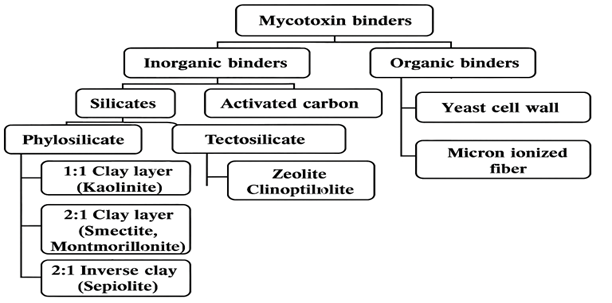
Figure 1. A diagram representing the classification of different mycotoxin binders by their source, nature and structural composition.
2.1. Inorganic binders
There is no consensus on the classification of clay binders that is acceptable to different disciplines such as agriculture, environment, or construction applications. Therefore, we report a classification of inorganic binders based on their properties to bind mycotoxins as proposed and updated by Murray (2007).
a) Silicate binders
Silicates are the most abundant elements found on earth crust (Kandel, 2018). Silicate is a mineral combining silicon dioxide (SiO2 4−) with a tetrahedral structure, where the silicon ion is in the center and surrounded by four oxygen atoms. The interaction of the positive silicon charges and negative oxygen charges results in an unbalanced structure. This allows the free oxygen charges to be bound to other silicon ions forming a chain of tetrahedral structures in different combinations, resulting in chains, sheets, rings, and three-dimensional structures. The tetrahedral sheet is the basis of silicate binders where different subgroups of silicate are formed in combination with other mineral ions in bi or three-dimensional structures. The two main subclasses of silicates are phyllosilicate (sheets of silicate) or tectosilicate (framework silicate, Figure 2).
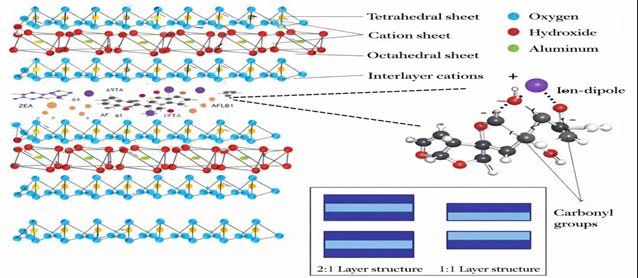
Figure 2. Molecular structure of octahedral and tetrahedral sheets of tectosilicate binders, and an illustration of the contribution of ions to the adsorption mechanism of mycotoxins.
b) Phyllosilicate binders
Phyllosilicates are bidimensional laminar or tubular minerals composed of tetrahedral silicate sheets linked to octahedral sheets of aluminum or magnesium hydroxides [(Al/MgOH)₆]. Charge imbalance within the octahedral layer, compensated by either two Al³⁺ or three Mg²⁺ ions, results in dioctahedral or trioctahedral structures. Based on layer stacking, phyllosilicates are classified as 1:1 types (e.g., kaolinite–serpentinite), consisting of one tetrahedral and one octahedral sheet, and 2:1 types (e.g., smectites), where an octahedral sheet is sandwiched between two tetrahedral sheets.
Isomorphic substitution of Si⁴⁺ or Al³⁺ with lower‑valence cations (e.g., Mg²⁺, Fe²⁺) generates negatively charged layers balanced by exchangeable interlayer cations, conferring swelling behavior and high cation‑exchange capacity essential for mycotoxin adsorption. Smectites, particularly montmorillonite, exhibit high adsorption efficiency, while bentonite, rich in montmorillonite, shows comparable binding properties (Murray, 2007).
c) Tectosilicate binders
Tectosilicate binders are crystalline aluminosilicate minerals, with zeolites as the main constituents. They are formed by three-dimensional assemblies of tetrahedral units linked through shared oxygen atoms, creating cage- or ring-like porous structures. These uniform pores contain exchangeable cations and water molecules, providing adsorption sites where potassium and calcium ions interact with mycotoxins depending on molecular size. Zeolites are classified based on crystal structure, chemical composition, cation type, pore size, and structural stability. Clinoptilolite is the most widely used zeolite due to its high resistance to low pH and elevated temperatures, functioning as a molecular sieve with pore sizes of approximately 3–8 Å. Thermal treatment or cation enrichment can further enhance its adsorption capacity (Eseceli et al., 2017).
d) Activated carbon
Activated carbon (AC) is an insoluble carbonaceous powder produced by pyrolysis of organic materials such as wood, bamboo, or coal at temperatures up to 2000 °C. An activation process is required to enhance its adsorption capacity by developing a highly porous structure. Chemical activation involves impregnation with agents such as KOH, H₃PO₄, or ZnCl₂ followed by heating, but often results in impurities and environmentally harmful residues. Physical activation uses oxidation with oxygen or CO₂ at 600–900 °C, producing highly microporous carbon with a large surface area (500–3000 m²/g). Adsorption efficiency is directly related to pore availability, with AC sources showing variable mycotoxin-binding capacity (Galvano et al., 1997).
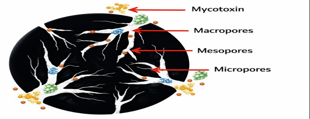
Figure 3. Structure of macro and micropores of activated carbon for the adsoprtion of mycotoxins and other nutrients.
2.2. Organic binders
a) Yeast Cell Wall (YCW)
The yeast cell wall (15–30% of yeast dry weight) is the main component responsible for mycotoxin adsorption. It consists of an inner layer rich in β-(1,3)- and β-(1,6)-D-glucans (50–60%), which provide structural rigidity and binding sites, linked to the membrane by chitin. Excess chitin reduces flexibility and mycotoxin affinity. The outer layer (≈40%) is composed of glucomannans and mannoproteins that determine surface properties. Mycotoxin-binding capacity increases with higher β-D-glucan content in the yeast strain (Jouany et al., 2005).
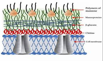
Figure 4. The composition of different yeast cell wall sheets and their components (adapted from Talavera et al., 2013).
b) Micro-ionized fiber
Micro‑ionized fibers are emerging mycotoxin binders (MTB) capable of adsorbing a wide range of mycotoxins. Various plant‑derived biomaterials, including grape pomace and stem, olive pomace, alfalfa hay, and wheat straw, have shown binding efficiencies ranging from 27 to 90%, depending on the material and mycotoxin type. Their adsorption relies on physico‑chemical interactions between mycotoxins and fiber components such as lignin, cellulose, and polyphenols, similar to silicate or activated carbon binders. However, high inclusion rates (≈20 kg/t) limit their use in monogastric diets, while ruminant diets may better tolerate them (Čolović et al., 2019).
3. Adsorption Mechanism of Different Binders
3.1. Mycotoxin binder properties
a) Silicate binders (clays and zeolites)
Silicate binders adsorb mycotoxins mainly through cation exchange capacity (CEC) and surface charge interactions, which are strongly influenced by pH and point of zero charge (PZC). At low pH, protonation reduces adsorption, whereas higher pH exposes negative charges that facilitate binding of cations interacting with mycotoxin carbonyl groups via weak ion‑dipole and Van der Waals force. Interlayer spacing is critical; sodium bentonite shows greater aflatoxin adsorption than calcium bentonite, while zeolites are limited by smaller pore size. Structural and organic modifications further enhance adsorption efficiency (Jaynes & Zartman, 2011).
b) Activated carbon (AC)
Activated carbon adsorbs mycotoxins primarily via hydrophobic interactions and π‑bonding, showing greater affinity for non‑polar toxins. Activation processes increase surface oxygen‑containing functional groups, enhancing polarity and enabling adsorption of polar mycotoxins such as aflatoxins and fumonisins. Adsorption efficiency is determined by surface area and pore size distribution, which must match mycotoxin molecular dimensions (Goto et al., 2015). The adsorption efficiency of activated carbon (AC) is strongly influenced by pore size and pore size distribution.
AC pores are classified into three categories: micropores (<2 nm), mesopores (2–50 nm), and macropores (>50 nm). Micropores contribute most to surface area and adsorption capacity, while meso- and macropores facilitate diffusion. If the pore size is not compatible with the molecular size of mycotoxins, diffusion into the pores is restricted. Limited accessibility to the internal pore surface reduces overall adsorption efficiency of AC.
c) Yeast cell wall (YCW)
Yeast cell wall binders act mainly through β‑ (1,3)‑D‑glucans, which interact with mycotoxins via Van der Waals forces between aromatic rings and glucan structures, as well as hydrogen bonding with hydroxyl, ketone, and lactone groups. Three‑dimensional conformational compatibility between the mycotoxin and glucan helices enhances complex stability and binding strength (Yiannikouris et al., 2004).
4. Mycotoxin properties
Physico-chemical characteristics of mycotoxins significantly influence the adsorption capacity of MTB (Galvano et al., 1997).
Classification of mycotoxins can be based on:
a) Polarity
Polarity reflects the charge distribution within a mycotoxin molecule. Mycotoxins can be classified as polar, non-polar, or intermediate. Aflatoxins (AF) and fumonisins (FUM) are the most polar mycotoxins. Zearalenone (ZEA) is non-polar. Deoxynivalenol (DON), T-2 toxin, and ochratoxin A (OTA) exhibit intermediate polarity.
b) Solubility
Solubility of mycotoxins in the surrounding medium is crucial for effective adsorption. Most mycotoxins are soluble in organic solvents such as methanol, acetonitrile, and acetone. Water solubility depends on polarity: More polar mycotoxins are generally more soluble in water.
c) Chemical structure, size, and shape
These structural features strongly affect the adsorption efficiency of mycotoxins. Aflatoxins (AF) are small, flat molecules, allowing easy entry into the interlayer spaces of binders, leading to higher adsorption. Fumonisins (FUM) have a large, branched molecular structure, which restricts their access to the interlayer space of MTB, resulting in reduced adsorption (Galvano et al., 1996).
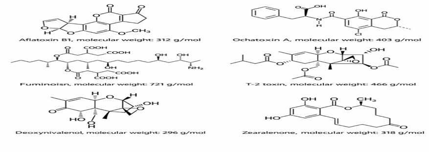
Figure 5. Chemical structure of the major mycotoxins and their molecular weight.
Table 1. Different types of Mycotoxins
| Name | Source | Types / Characteristics | Target organ | Harmful effects |
| Aflatoxin | Aspergillus flavus, Aspergillus parasiticus | Naturally occurring aflatoxins include B1, B2, G1, and G2; among these, B1 is found in the highest concentration and is the most toxic | Liver, Kidney, Spleen, Testes, Thymus | Loss of egg production, anaemia, haemorrhage, liver damage, paralysis, poor FCR, immunosuppression |
| Ochratoxin | Aspergillus ochraceus, Penicillium verridicatum | Four types: A, B, C, and D; Ochratoxin A is the most toxic | Kidney, Liver, Thymus | Gout, immunosuppression, reduced weight gain, reduced egg production and egg weight |
| Trichothecene (T-2 toxin) | Fusarium spp. | — | — | Reduced feed intake, decreased growth, immunosuppression |
| Citrinin | Penicillium spp. | — | Kidney | Wet droppings, reduced weight gain |
| Oosporein | — | — | Kidney | Gout, high mortality |
| Moniliformin | Fusarium moniliforme | — | — | Reduced feed intake and body weight; in chicks causes heart damage and death; in layers reduces rate of laying and delays peak production |
| Fumonisins | Fusarium moniliforme | — | — | More harmful to pigs and horses; poultry are more resistant |
| Zearalenone | — | — | — | — |
| Ergotism | — | — | — | Reduced feed intake and growth; death of tissues of beak, comb, and toes; diarrhoea; vesicles and crusts on comb and wattle; reduced egg production |
| Fusarochromanone | — | — | — | Leg deformities in chicks |
Table 2. Summary of studies that determine the capacity of different mycotoxin binders to adsorb nutrients
| Binder | Nutrient interaction effects | Observation | Reference |
| Bentonite | High adsorption of vitamins E, B1, B2, and B6, and amino acids (lysine, methionine, threonine); low adsorption of vitamins A, D, and B3 | In vitro simulation of gastrointestinal tract | Kihal et al. (2020, 2021) |
| High adsorption of vitamin B1 and pepsin; no adsorption of vitamins D and E | In vitro gastric fluid simulation | Barrientos-Velázquez et al. (2016) | |
| High adsorption of vitamins B12 and B8; no adsorption of vitamin B5 | In vitro gastric fluid simulation and real gastric fluid | Vekiru et al. (2007) | |
| High adsorption of vitamin B6; adsorption of Zn and Co; no adsorption of Cu and Mn | In vitro aqueous solution | Tomasevic-Canovic et al. (2000) | |
| Adsorption of vitamin B2 | In vitro aqueous solution | Mortland and Lawless (1983) | |
| No adsorption of vitamin A | In vivo in chicks | Pimpukdee et al. (2004) | |
| No adsorption of vitamin A | In vivo in chicks | Afriyie-Gyawu (2004) | |
| MMT | High adsorption of vitamins E, B1, B2, and B6, and amino acids (lysine, methionine, threonine); low adsorption of vitamins A, D, and B3 | In vitro simulation of gastrointestinal tract | Kihal et al. (2020, 2021) |
| Adsorption of vitamin B1 | In vitro gastric fluid simulation | Ghanshyam et al. (2009) | |
| Adsorption of protein, urea, and antibiotics | In vitro agar culture | Pinc (1962) | |
| No adsorption of vitamins A, D, E, B1, and B6 | In vivo in dairy cows | Kihal et al. (2022) | |
| No adsorption of vitamins A and B1 | In vivo in dairy cows | Maki et al. (2016) | |
| Ca-MMT | High adsorption of vitamins E, B1, B2, and B6, and amino acids (lysine, methionine, threonine); low adsorption of vitamins A, D, and B3 | In vitro simulation of gastrointestinal tract | Kihal et al. (2020, 2021) |
| Adsorption of vitamins B8 and B12 | In vitro simulation of gastric fluid and real gastric fluid | Vekiru et al. (2007) | |
| Clinoptilolite | High adsorption of vitamins E, B1, B2, and B6, and amino acids (lysine, methionine, threonine) | In vitro simulation of gastrointestinal tract | Kihal et al. (2020, 2021) |
| No adsorption of vitamins A, D, and B3; no adsorption of amino acids (tryptophan, phenylalanine) | In vitro aqueous solution | Tomasevic-Canovic et al. (2000) | |
| HSCAS | No adsorption of vitamins A, B1, and minerals (Zn, Mn) | In vivo in chicks | Chung et al. (1990) |
| Sepiolite | High adsorption of vitamins E, B1, B2, and B6, and amino acids (lysine, methionine, threonine); low adsorption of vitamins A, D, and B3 | In vitro simulation of gastrointestinal tract | Kihal et al. (2020, 2021) |
| Zeolite | High adsorption of vitamins E, B1, B2, and B6, and amino acids (lysine, methionine, threonine); low adsorption of vitamins A, D, and B3 | In vitro simulation of gastrointestinal tract | Kihal et al. (2020, 2021) |
¹MMT, montmorillonite.
²AC, activated carbon.
³HSCAS, hydrated sodium calcium aluminosilicate.
Table 3. Regulatory guidance levels for major mycotoxins in finished poultry feed as established by the European Union (EU) and United States Food and Drug Administration (FDA). Values are expressed in mg/kg diet. EU limits in finished feed set according to the European Commission Recommendation 2006/576/EC and the European Commission Directive 2003/100/EC; USA limits in finished feed set according to the Food and Drug Administration regulatory guidance for toxins and contaminants.
| Guidance | Deoxynivalenol (DON) (mg/kg) | Fumonisins (FUM) (mg/kg) | Zearalenone (ZEA) (mg/kg) | Aflatoxin (AF) (mg/kg) | Ochratoxin A (OTA) (mg/kg) | T-2 Toxin (mg/kg) |
| EU limits (EC guidance) ¹ | 5 | 20 | – | 0.02 | 0.1 | No guidance level established |
| USA limits (FDA guidance) ² | 5 | 50 | No guidance level established | 0.1 | No guidance level established | No guidance level established |
Table 4. Ranges of ratios of mycotoxin binder to mycotoxins doses used in in vitro tests to determine the mycotoxin adsorption capacity of different mycotoxin binders
| Binders | AFB1 (mg: µg) | DON (mg: µg) | FUM (mg: µg) | OTA (mg: µg) | T-2 (mg: µg) | ZEA (mg: µg) |
| AC | 1:0.008–1:461 | 1:0.2–1:90 | 1:0.92–1:25 | 1:0.025–1:125 | 1:0.1 | 1:0.05–1:20 |
| Bentonite | 1:0.0007–1:200 | 1:0.2–1:12 | 1:2–1:20 | 1:0.002–1:12.5 | 1:0.1–1:0.2 | 1:0.1–1:20 |
| Clinoptilolite | 1:0.2–1:40 | 1:1.2 | – | 1:2 | – | 1:0.05–1:12 |
| HSCAS | 1:0.002–1:600 | 1:0.4–1:12 | 1:2–1:20 | 1:0.025–1:10 | 1:0.1 | 1:0.001–1:20 |
| MMT | 1:0.002–1:20 | 1:0.5–1:12 | 1:2.5 | 1:0.025 | 1:0.1 | 1:0.05–1 |
| Sepiolite | 1:1.6–1:10 | 1:2–1:12 | 1:2 | 1:10 | – | 1:0.5 |
| YCW | 1:0.001–1:461 | 1:0.2–1:12 | 1:2–1:20 | 1:0.001–1:10 | 1:0.1 | 1:0.001–1:20 |
| Zeolite | 1:0.002–1:20 | 1:0.2–1:10 | 1:0.2–1:20 | 1:0.016–1:5 | 1:0.2 | 1:0.05–1:20 |
1AC, activated carbon; HSCAS, hydrated sodium calcium aluminosilicate; MMT, montmorillonite; YCW, yeast cell wall. 2AFB1, aflatoxin B1; DON, deoxynivalenol; FUM, Fumonisin; OTA, ochratoxin; T-2, T-2 toxin; ZEA, zearalenone.
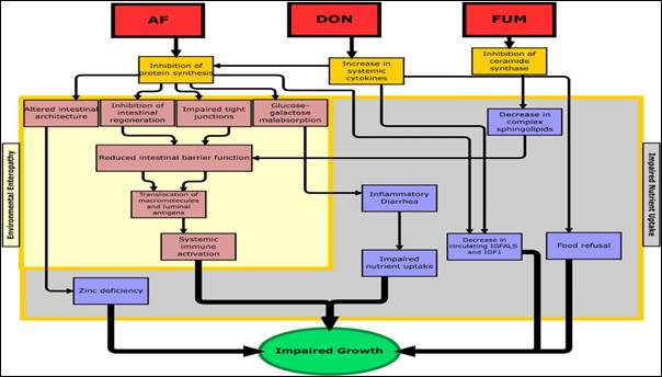
Figure 6. A conceptual framework for the effect of mycotoxin exposure on growth retardation (Smith et.al.2012).
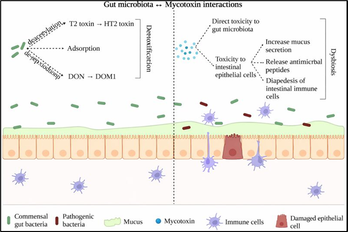
Fig. 7. Gut microbiota and mycotoxins interactions. Illustration of the bidirectional interactions between gut microbiota and dietary mycotoxins in poultry. Mycotoxins can disrupt microbial community structure, reduce beneficial populations, and impair microbially derived functions, including short-chain fatty acid (SCFA) production and mucosal barrier maintenance. Conversely, the gut microbiota can biotransform certain mycotoxins into less toxic metabolites or modulate host responses to exposure. Disruption of this balance may compromise gut integrity, immunity, and overall performance.
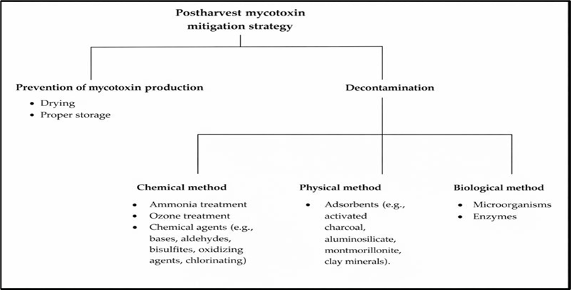
Figure 8. Diagrammatic representation for postharvest mycotoxin mitigation strategies in broiler and layer chickens’ production.
Stallen offers a comprehensive range of world‑class mycotoxin binders, including D‑Tox and Alusil MOS Plus, both of which provide broad‑spectrum protection against diverse mycotoxins commonly found in poultry feed.
5.2. The characteristic features of D-Tox compare with other common toxin binder.
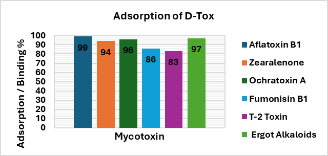
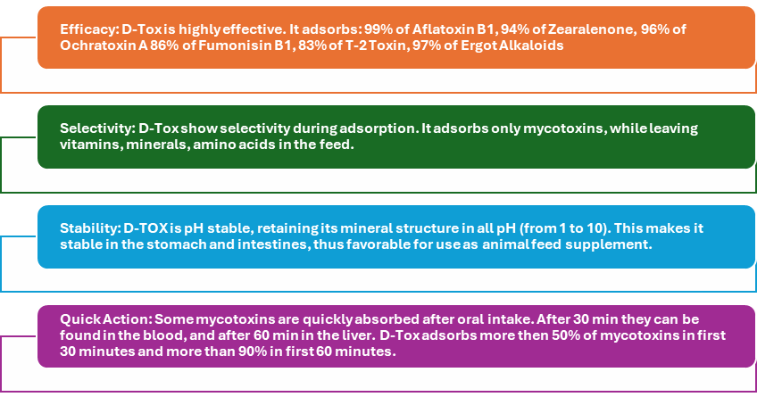
5.3. D-Tox Benefits:
D‑Tox provides effective and comprehensive control of all major categories of mycotoxins, thereby improving performance and productivity in poultry.
6. Alusil MOS Plus
Alusil MOS Plus contains HSCAS (Activated Hydrated Sodium Calcium Aluminosilicates), activated charcoal, MOS (Mannan Oligosaccharides), copper oxinate, organic acids (propionic, benzoic, acetic, and sorbic acids), lipotropic agents, and spirulina.
6.1. Mechanism of action of Alusil MOS Plus
HSCAS acts as an enterosorbent that tightly and selectively binds aflatoxins in the GI tract of animals decreasing their bioavailability and associated toxicity. Mannan Oligosaccharides act as bio-binder which helps absorption of pathogens, improves intestinal function & immune modulation. Organic acids kill harmful bacteria and fungi. Activated charcoal which is 200 MT grade helps to bind pesticides and toxins like ochratoxin. Copper oxinate is broad spectrum anti-fungal agent which acts against spp. of Aspergillus, Fusarium, Penicillium, Candida etc. Lipotropic agent and herbal ingredients help in mobilizing fat which are accumulated in liver due to damage caused by toxins. Spirulina helps in restoring the liver damaged by toxins.
6.2. The characteristic features of Alusil MOS Plus over other toxin binder.
| Component | Function |
| HSCAS | Enters bloodstream and binds to aflatoxins. |
| Activated Charcoal | Binds multiple mycotoxins including aflatoxins, ochratoxins A, zearalenone, and along with bacterial endotoxins & pesticides in the intestinal tract, preventing their reabsorption. |
| MOS | Binds to pathogenic bacterial fimbriae, increases mucin secretion & acts as a bactericidal agent. |
| Copper Oxinate | Acts as antifungal agent by inhibiting mould growth. |
| Organic Acids | Acetic, benzoic, propionic & sorbic acids inhibit fungal growth and reduce aflatoxin secretion. |
| Lipotropic Agent | Aids in mobilising fat accumulated in liver due to toxin damage. |
| Spirulina | Helps restore liver function damaged by toxins. |
6.3. Alusil MOS Plus Benefits:
Alusil MOS Plus acts as a broad-spectrum mould inhibitor and supports bio-neutralization of mycotoxins, helping prevent toxin-related damage and overcome clinical symptoms of aflatoxicosis. It protects the immune system, enhances vaccine and drug response, improves pellet quality, functions as an anti-caking agent in feed, and does not bind essential vitamins and minerals.
7. Conclusion
Stallen South Asia Pvt. Ltd. offers effective toxin binders, D-tox and Alusil MOS Plus, which are helpful in the detoxification of mycotoxins in poultry. D-tox provides broad‑spectrum control of both polar and non‑polar mycotoxins through pH‑stable adsorption and pre‑absorptive detoxification, without binding amino acids, fat‑soluble vitamins, minerals, or micronutrients. It also sequesters heavy metals, endotoxins, and biogenic amines, reducing immunosuppression. Alusil MOS Plus alleviates aflatoxicosis, protects immune function, enhances vaccine and drug response, and improves feed quality through anticaking and pellet‑stabilizing effects.
References
Agboola, A.F., Omidiwura, B.R.O., Odu, O., Odupitan, F.T., Iyayi, E.A., 2015. Effect of probiotic and toxin binder on performance, intestinal microbiota and gut morphology in broiler chickens. Journal of Animal Science Advances 5(7), 1369. https://doi.org/10.5455/jasa.20150709085312.
Barrientos-Velazquez, A.L., Arteaga, S., Dixon, J.B., Deng, Y., 2016. The effect of pH, pepsin, exchange cation, and vitamins on aflatoxin adsorption on smectite in simulated gastric fluids. Applied Clay Science 120, 17–23. https://doi.org/10.1016/j.clay.2015.11.014.
Čolović, R., Puvača, N., Cheli, F., Avantaggiato, G., Greco, D., Đuragić, O., Pinotti, L., 2019. Decontamination of mycotoxin-contaminated feedstuffs and compound feed. Toxins 11, 617. https://doi.org/10.3390/toxins11110617.
Choudhary, A.K., Kumari, P., 2010. Management of mycotoxin contamination in preharvest and post-harvest crops: present status and future prospects. Journal of Phytology 2.
Desjardins, A., Plattner, R., Shackelford, D., Leslie, J., Nelson, P., 1992. Heritability of fumonisin B1 production in Gibberella fujikuroi mating population A. Applied and Environmental Microbiology 58, 2799–2805.
Di Gregorio, M.C., Neef, D.V., Jager, A.V., Corassin, C.H., Carao, A.C.P., Albuquerque, R., Azevedo, A.C., Oliveira, C.A.F., 2014. Mineral adsorbents for prevention of mycotoxins in animal feeds. Toxin Reviews 33, 125–135. https://doi.org/10.3109/15569543.2014.905604.
Eseceli, H., Ozcan, M.A., Demir, E., Bilal, T., 2017. Increasing the adsorption of clinoptilolite as toxin binder by heat treatment. Bulgarian Journal of Veterinary Medicine 20, 18–26.
European Commission (EC), 2006. EC regulation No 401/2006 laying down the methods of sampling and analysis for the official control of the levels of mycotoxins in foodstuffs. Official Journal of the European Communities L 70, 12–34.
Food and Drug Administration (FDA), 2001. Guidance for Industry: Fumonisin levels in human foods and animal feeds; Final guidance. US FDA, Silver Spring, MD, USA.
Galvano, F., Pietri, A., Bertuzzi, T., Bognanno, M., Chies, L., Angelis, A., Galvano, M., 1997. Activated carbons: in vitro affinity for fumonisin B1 and relation of adsorption ability to physicochemical parameters. Journal of Food Protection 60, 985–991. https://doi.org/10.4315/0362-028X-60.8.985.
Gimeno, A., Martins, M.L., 2007. Micotoxinas y micotoxicosis en animales y humanos. 1st ed. Miami, FL, USA: Special Nutrients; p. 128.
Goto, T., Amano, Y., Machida, M., Imazeki, F., 2015. Effect of polarity of activated carbon surface, solvent and adsorbate on adsorption of aromatic compounds from liquid phase. Chemical and Pharmaceutical Bulletin 63, 726–730. https://doi.org/10.1248/cpb.c15-00039.
Greco, M.V., Franchi, M.L., Rico Golba, S.L., Pardo, A.G., Pose, G.N., 2014. Mycotoxins and mycotoxigenic fungi in poultry feed for food-producing animals. The Scientific World Journal, 1–10. https://doi.org/10.1155/2014/968215.
Gruber-Dorninger, C., Jenkins, T., Schatzmayr, G., 2019. Global mycotoxin occurrence in feed: a ten-year survey. Toxins 11, 375. https://doi.org/10.3390/toxins11070375.
Jaynes, W.F., Zartman, R.E., 2011. Aflatoxin toxicity reduction in feed by enhanced binding to surface-modified clay additives. Toxins 3, 551–565. https://doi.org/10.3390/toxins3060551.
Jouany, J.P., Yiannikouris, A., Bertin, G., 2005. How yeast cell wall components can alleviate mycotoxicosis in animal production and improve the safety of edible animal products. Journal of Animal Science 14, 171–190. https://doi.org/10.22358/jafs/70361/2005.
Kandel, R., 2018. Potential for using aluminosilicates for removal of heavy metals and mycotoxins from feed and water [Master’s thesis]. Ås, Norway: Norwegian University of Life Sciences.
Mottet, A., Tempio, G., 2017. Global poultry production: current state and future outlook and challenges. World’s Poultry Science Journal 73, 245–256.
Murray, H.H., 2007. Developments in clay science: Occurrences, processing and applications of kaolins, bentonites, palygorskite–sepiolite, and common clays, Vol. 2. UK: Elsevier Science & Technology; pp. 1–180.
Smith, L., Stoltzfus, R., Prendergast, A., 2012. Food chain mycotoxin exposure, gut health, and impaired growth: A conceptual framework. Advances in Nutrition 3, 526–531. https://doi.org/10.3945/an.112.002188.
Weaver, A.C., Weaver, D.M., Adams, N., Yiannikouris, A., 2021. Co-occurrence of 35 mycotoxins: a seven-year survey of corn grain and corn silage in the United States. Toxins 13, 516.
Yiannikouris, A., Francois, J., Poughon, L., Dussap, C.G., Bertin, G., Jeminet, G., Jouany, J.P., 2004. Adsorption of zearalenone by β-D-glucans in the Saccharomyces cerevisiae cell wall. Journal of Food Protection 67, 1195–1200. https://doi.org/10.4315/0362-028X-67.6.1195.
18th February 2026, Patna, Bihar.
To address the evolving disease landscape in poultry production, Stallen South Asia Pvt. Ltd. recently organized a focused technical seminar in Patna, Bihar, attended by 30 broiler breeder and layer farmers from the region.
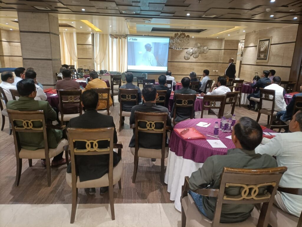
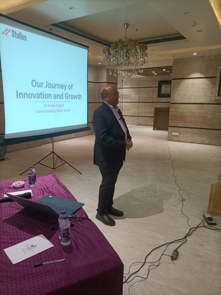
The keynote session was delivered by Dr. Sushil Dhariwal, who spoke on “Emerging Challenges in IB, Mycoplasma and Marek’s Control in Poultry Production.” He highlighted the increasing complexity of Infectious Bronchitis due to variant strains, the persistent economic impact of Mycoplasma (MG & MS) infections, and the growing importance of strong early immunization strategies against Marek’s disease. Emphasis was placed on structured vaccination programs supported by monitoring, biosecurity, and scientific field evaluation.
Following his keynote address, farmers actively participated in an engaging and practical Q&A session with Dr. Dhariwal. The discussion covered field-level challenges such as fluctuating ELISA titres during lay, nephropathogenic IB concerns, Mycoplasma persistence in breeder flocks, and early-age Marek’s-related mortality patterns. The interactive exchange reflected the keen interest of producers in strengthening preventive health strategies through scientific understanding.


The program commenced with a welcome address by Mr. Biplab Deb, Regional Manager – East. An overview of the company’s philosophy and legacy was presented by Dr. Sanjay Singhal, COO, who spoke about the origin and growth of Stallen, its evolution as a science-driven animal health organization, and its unwavering commitment to quality and ethical practices. He underlined that the company’s foundation is built on technical excellence, stringent quality standards, and long-term partnerships with poultry producers. Dr. Singhal also highlighted that Stallen imports high-quality live and killed vaccines in collaboration with Fatro S.p.A., Italy, ensuring access to internationally benchmarked vaccine technology for the Indian poultry sector. The portfolio discussed during the seminar included vaccines for Marek’s disease, Mycoplasma (MG & MS), and IB–ND combinations, with emphasis on their strategic integration into breeder and layer vaccination programs.
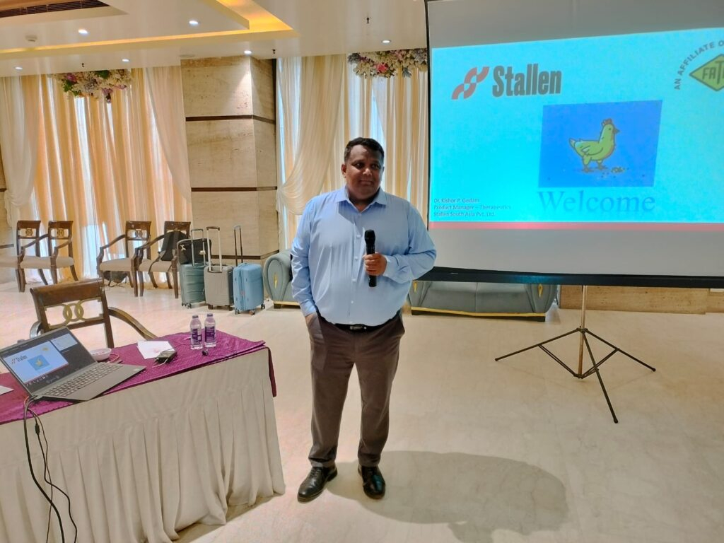

The seminar concluded with a vote of thanks by Mr. Mmukesh Singh, Area Manager, acknowledging the enthusiastic participation of farmers and the valuable scientific insights shared during the session.
Such knowledge-sharing initiatives continue to play a crucial role in reinforcing disease preparedness and promoting sustainable productivity in Eastern India’s poultry industry.
Dr. Amit Janbandhu, Dr. Sanjay Singhal
1. Introduction
Necrotic enteritis (NE) is a significant burden on the poultry industry, causing gut damage that reduces nutrient utilization and productivity. Production losses due to NE and current control methods are estimated to cost the global broiler industry approximately USD 6 billion annually (Wade and Keyburn, 2015). These losses are associated with decreased production performance, mortality of up to 1% per day, treatment costs, and carcass condemnation due to cholangiohepatitis (Immerseel,et.al, 2004 & Timbermont, et.al,2011). Clinical and subclinical forms of NE have been recognized (Van Immerseel et al., 2004); the clinical form results in acute disease and bird mortality. Although flock health and productivity can be maintained using IFA-free practices (Parent et al., 2020), strong industry moves away from IFAs have increased the need for alternative approaches. Consequently, a wide range of feed additives and treatments have been investigated and commercialized to control NE.
NE is caused by Clostridium perfringens and primarily affects chickens from 2 weeks to 6 months of age. In humans, C. perfringens intoxications are the third most common bacterial foodborne disease after Salmonella and Campylobacter, with 359–2173 cases reported annually in the United States. Poultry and poultry products account for 30% of outbreaks, and 92% are traced to meat and poultry as a single identified food commodity (Grass et.al, 2013). Research into antibiotic alternatives that improve gut health and immune status has intensified; however, current alternatives are less effective than antibiotics in controlling NE. A greater understanding of C. perfringens virulence factors, NE pathogenesis, and host responses is required to develop effective control strategies and new supplements, as these aspects are not yet fully understood and remain under investigation.
2. Necrotic Enteritis
Etiology: Necrotic enteritis is caused by Clostridium perfringens, a Gram-positive, rod-shaped, anaerobic bacterium that forms oval subterminal spores. Unlike most clostridia, C. perfringens consists of relatively large, encapsulated, non-motile rods (0.6–2.4 × 1.3–9.0 µm). Colonies are smooth, round, and glistening, with an inner zone of complete hemolysis mediated by theta-toxin and an outer zone of incomplete hemolysis caused by alpha-toxin (Cato et al., 2002).
Clostridium perfringens is classified into five biotypes (A–E) based on the production of four major lethal toxins: alpha, beta, epsilon, and iota. In addition, enterotoxin (CPE) and beta2 (CPB2) toxins are considered important in enteric diseases; however, their roles in avian C. perfringens–associated enteric disease remain unclear (Crespo et al., 2007). All five types produce toxins: type A (α), type B (α, β, ε), type C (α, β), type D (α, ε), and type E (α, ι). Necrotic enteritis is primarily associated with C. perfringens types A and C caused by α and net B toxins (Fisher et al,2005)., with infections in poultry mainly caused by type A and, to a lesser extent, type C. Because type A is highly prevalent in the intestines of healthy birds, its pathogenic role remains controversial (Smedley et.al,2004). Moreover, strains isolated from NE outbreaks have not been shown to produce higher levels of alpha toxin than isolates from clinically healthy broilers.
Fig 1. Microscopic appearance of Clostridium Perfringes
Table 1: The most important C. perfringens toxins
| Toxin | Gene location | Biological activity |
| Alpha toxin | Chromosome | Cytolytic, haemolytic, dermonecrotic, Lethal |
| Beta toxin | Plasmid | Cytolytic, dermonecrotic, lethal |
| Epsilon toxin | Plasmid | Oedema in various organs: liver, kidney and central nervous system |
| Iota toxin | Plasmid | Disruption of actin cytoskeleton and cell barrier integrity |
| Beta2 toxin | Plasmid | Cytolytic, lethal |
| Enterotoxin | Chromosome/ Plasmid | Cytotoxic, lethal, causes diarrhea by leakage of water and ions |
| Theta toxin | Chromosome | Lyses red blood cells and modulates the host inflammatory response |
(Source: Wise and Siragusa et.al, 2005)
3. Epidemiology
3.1. Source of Infection and Transmission: Clostridium perfringens is a naturally occurring bacterium in poultry production environments. It is present in dust, soil, feces, feed, poultry litter, eggshell fragments, fluff, and the intestinal tract of poultry. Feces of wild birds may also contain elevated numbers of C. perfringens, further introducing the organism into poultry facilities. Environmental sampling on poultry farms detected C. perfringens on wall swabs (53%), fan swabs (46%), fly strips (43%), dirt outside entrances (43%), and boot swabs (29%), demonstrating its ubiquitous environmental presence (Craven et.al,2001). Transmission occurs primarily via the fecal–oral route and through contaminated feed, water, housing structures, insects, and direct contact between infected and susceptible birds. During necrotic enteritis (NE) outbreaks, contaminated feed or litter are considered major sources, and contaminated feed components have also been implicated. Vertical transmission is possible, as C. perfringens has been found in the yolk sac of embryonated eggs, suggesting transmission within integrated broiler operations (Craven et.al, 2003). Additionally, C. perfringens may be transmitted mechanically and/or biologically by house flies in poultry houses, contributing to NE development (Dhillon et.al,2004).
3.2. Predisposing factors
Necrotic enteritis (NE) develops when one or more predisposing factors are present, particularly intestinal mucosal damage. Damage caused by coccidial pathogens releases growth factors that promote C. perfringens proliferation in the intestinal lumen. Broilers inoculated with Eimeria spp. and fed C. perfringens–contaminated feed show higher mortality than birds fed contaminated feed alone. Physical damage from litter eating or fibrous diets may also alter the mucosa (Williams et.al,2005).
Management factors such as feeding practices, water supply, temperature control, and ventilation contribute to NE. Delayed initial feeding impairs gut-associated lymphoid tissue development. Nutritional stress from unbalanced diets, especially low energy-to-protein ratios, increases feed intake, nitrogen levels in digesta, and susceptibility to clostridial overgrowth. Diet composition strongly influences NE. Diets rich in wheat, rye, and barley contain indigestible non-starch polysaccharides that increase digesta viscosity, slow gut transit, and favor anaerobic bacteria. Broilers fed wheat-, rye-, or barley-based contaminated diets have higher mortality than those fed corn-based diets. Pelleted diets reduce intestinal C. perfringens, whereas high-protein diets (e.g., fishmeal) and bone meal increase NE risk (Kocher et.al,2003).
Immunosuppression increases NE susceptibility. Use of Infectious bursal disease (IBD) vaccines has been associated with increased NE lesion severity, even at normal doses. Stressful conditions may further predispose birds to NE, but immunosuppression is inappropriate when evaluating vaccines (Nikpiran et.al,2008).
4. Clinical Signs and Lesions
4.1. Clinical signs:
Subclinical necrotic enteritis (SNE) shows no obvious clinical signs and is usually detected under field conditions at processing plants through carcass rejection. SNE may be suspected based on reduced weight gain, poor feed conversion efficiency, increased moisture in droppings, and wet litter, most commonly at 2–5 weeks of age without increased mortality.
Clinical necrotic enteritis (NE) typically affects broiler chicks between 2–6 weeks of age and presents with sudden onset of diarrhoea and intestinal mucosal necrosis. Affected birds are depressed, anorectic, have ruffled feathers, and tend to huddle. In advanced stages, birds become laterally recumbent, immobile, and die rapidly. The disease course is usually short, and birds are often found dead without prior clinical signs. Acute signs include severe depression, reduced appetite, reluctance to move, ruffled feathers, and diarrhoea, with illness lasting only 1–2 hours [20]. Mortality in affected flocks may range from 1% to 50%.
Birds that die of NE have a foetid odor, dehydration, dark and dry pectoral muscles, and pale kidneys. The unopened intestine is darker than normal and distended due to bile-stained contents (Long, et.al,2007)
4.2. Gross lesions:
Lesions of SNE are characterized by necrotic lesions in the intestinal wall and liver, occurring in one or more intestinal regions. Mild lesions appear as small ulcers or light-yellow spots on the mucosal surface, mainly in the jejunum and ileum and less commonly in the caeca. Severe lesions may involve membranes covering large intestinal segments, including the colorectal region and caecal tonsils. Liver abnormalities, primarily enlargement and occasional congestion, are also reported.
Clinical NE is characterized by extensive mucosal necrosis of the small intestine, covered with a yellow-brown or bile-stained pseudomembrane. Gross lesions are mainly confined to the small intestine but may also involve the liver and kidneys. At necropsy, the duodenum, jejunum, and ileum are thin-walled, friable, gas-filled, and distended with dark brown fluid. Ulcers may occur singly or in aggregates. In severe cases, a fibrino-necrotic or diphtheritic membrane covers large segments, often involving two-thirds of the jejunum and ileum (McDevitt et.al,2006).
4.3. Histopathological changes:
Microscopically, NE lesions consist of coagulative necrosis at the villous apices with a clear demarcation between necrotic and viable tissue. Degeneration may extend into the submucosa. Regeneration is characterized by epithelial proliferation, connective tissue formation, and reduced goblet, columnar, and epithelial cells, resulting in short, flattened villi with reduced absorptive surface. The pseudomembrane consists of necrotic villi, inflammatory cells, and bacterial aggregates (Olkowski et.al.2008).

Fig. 2. Gross pathological changes in NE: A. Jejunum of a bird showing ballooning. B. Jejunum showing small blackish necrotic spots. C. Jejunum showing Turkish towel appearance. D. Duodenum showing congestion.
5. Pathogenesis
Pathogenesis describes the complex and dynamic host–pathogen interactions at the molecular level and is essential for developing effective control measures. Bacterial pathogenesis involves six overlapping phases: colonization, growth and proliferation, nutrient acquisition, evasion of host defenses, host tissue injury, and transmission. In rapidly growing pathogens such as Clostridium perfringens, these phases occur almost simultaneously.
Colonization requires degradation of the intestinal mucus layer, which normally acts as a physical barrier. Intestinal mucins provide binding sites for bacterial adhesins, and pathogenic C. perfringens secrete bacteriocins (perfrins) that displace commensal Clostridium species. The organism produces glycoside hydrolases and chitinases that degrade mucins, providing nutrients and enabling microcolony formation on the mucosa. Predisposing factors such as Eimeria infection stimulate inflammation, increased mucin production, and release of essential amino acids, all of which enhance C. perfringens growth and colonization (Collier et.al,2008).
Once a threshold density is reached, an Agr-like quorum-sensing system activates the VirS–VirR regulatory system and virulence genes, including NELoc-1 involved in adhesion. Degradation of the mucus layer allows pore-forming toxins to access epithelial cells. Proteolytic and collagenolytic enzymes damage the epithelium, disrupt intercellular junctions, spread through the lamina propria, and cause epithelial necrosis and sloughing. Quorum-sensing–regulated secretion of alpha toxin and perfringolysin promotes biofilm formation on the exposed submucosa, enhancing bacterial persistence and protection from host immunity and antibiotics.
Intestinal integrity depends on tight junctions, particularly claudin-3 and claudin-4, which serve as receptors for C. perfringens enterotoxin (CPE). CPE binding forms small complexes that oligomerize into large CH1 complexes, leading to pore formation in the cell membrane. These pores allow calcium influx, resulting in epithelial cell death (Chakrabarti, et.al,2005).
Gross lesions of necrotic enteritis primarily affect the jejunum and ileum, with occasional involvement of the duodenum and ceca. The intestine is thin, friable, gas-distended, and covered by a tan orange pseudomembrane, producing the characteristic “dirty Turkish towel” appearance. Pseudomembrane formation is most common in the jejunum. Subclinical necrotic enteritis is associated with hepatitis or cholangiohepatitis and gall bladder distension with flocculent material. Bile acids promote sporulation and enterotoxin production by C. perfringens, explaining the higher lesion frequency in the upper small intestine, particularly the duodenum and jejunum (Park et.al,2018).

Fig.3. Pathogenesis of necrotic enteritis in broiler chickens causes destruction of epithelial cells of intestine that leads to blood-stained diarrhea.
6. Feed Additives Used to Control Necrotic Enteritis
A wide range of feed additives has been studied for their effects on necrotic enteritis (NE). Most commercial additives do not target Clostridium perfringens directly but improve gut health, microbiota balance, and immune competence, often with overlapping and interconnected effects (Granstad et al., 2020). Short-chain fatty acids, particularly butyrate, provide energy to enterocytes and support beneficial microbiota. Butyrate can be supplied directly in protected form or indirectly via prebiotics, probiotics, phytobiotics, or postbiotics (Liu et al., 2021). Combinations of additives, such as fatty acids with phytobiotics, are often more effective.
Probiotics, including single or multi-strain bacteria, yeasts, or cultured cecal microbiota, can directly inhibit C. perfringens, compete for gut niches, improve microbiota composition, gut integrity, or immune function. Prebiotics, fatty acids, and phytobiotics have also been reviewed for general health and NE-specific applications (Gomez-Osorio et al., 2021).
Novel approaches include bacteriophages and their endolysins, which specifically target C. perfringens, and bacteriocins produced by bacteria for antimicrobial effects. Passive immunization using egg yolk antibodies or engineered single-chain antibodies has shown potential in reducing NE ( Gangaiah et al., 2022).
Commercial feed supplements such as StalBMD (BMD), Magnox (lincomycin), and Stalgro (enramycin) by Stallen South Asia Pvt. Ltd are also used to prevent NE caused by C. perfringens.
7) Mechanism of Action
7.1) Bacitracin Methylene Disalicylate (BMD)
Bacitracin Methylene Disalicylate (BMD) is a polypeptide antibiotic used to prevent and treat Gram-positive bacterial infections. It inhibits bacterial cell wall synthesis by blocking dephosphorylation of bactoprenol phosphate, preventing peptidoglycan transport. This disrupts cell wall formation, causing bacterial lysis. BMD is bactericidal, particularly against actively dividing Staphylococcus and Streptococcus species.
Fig.4. Bactericidal effect on growing bacteria.

7.2) Lincomycin Hydrochloride
Lincomycin hydrochloride, a lincosamide antibiotic derived from Streptomyces lincolnensis, is used against Gram-positive and some anaerobic bacteria. Its primary mechanism is inhibition of bacterial protein synthesis by binding to the 50S ribosomal subunit, specifically at the peptidyl transferase center. This blocks the ribosomal exit tunnel, preventing elongation of polypeptide chains and halting protein synthesis, which stops bacterial growth and replication. Lincomycin may also have a secondary effect on bacterial cell wall synthesis, enhancing its overall antibacterial activity.
Fig 5. Protein synthesis inhibition within bacterial cells.
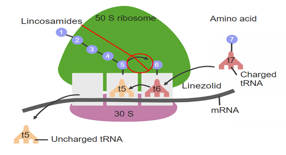
7.3. Enramycin
Enramycin acts as an inhibitor of the enzyme (MurG), which is essntial for wall biosynthesis in gram +ve bacteria. MurG catalyzes the tranglycosylation reaction in the last step of peptidoglycan biosynthesis. Hence inhibiting this step greatly compromises cell wall integrity leading to cell lysis.
Figure 6. Membrane steps of the bacterial peptidoglycan synthesis pathway.
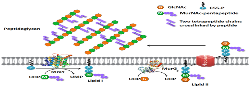
References
Experimental induction of necrotic enteritis inbroiler chickens by Clostridium perfringens isolates from the outbreaks in Iran. Journal of Veterinary Research,2008, 63: 127-132.
Cato, P., L. George and M. Finegold, 2002. Genus c l o s t r i d i u m . B e r g e y ‘ s m a n u a l o f systematicbacteriology, USA., 2: 1141-1200.
Chakrabarti, G.; McClane, B.A. The importance of calcium influx, calpain and calmodulin for the activation of CaCo-2 cell death pathways by Clostridium perfringens enterotoxin. Cell. Microbiol. 2005, 7, 129–146. [PubMed].
Collier, C.T.; Hofacre, C.L.; Payne, A.M.; Anderson, D.B.; Kaiser, P.; Mackie, R.I.; Gaskins, H.R. Coccidia-induced mucogenesis promotes the onset of necrotic enteritis by supporting Clostridium perfringens growth. Vet. Immunol. Immunopathol. 2008, 122, 104–115. [CrossRef].
Craven, E., A. Cox, S. Bailey and E. Cosby, 2003. Incidence and tracking of Clostridium perfringensthrough an integrated broiler chicken operation.Avian Diseases, 47(3): 707-711.
Craven, E., J. Stern, S. Bailey and A. Cox, 2001. Incidence of Clostridium Perfringens in broiler chickens and their environment during production and Processing. Avian Diseases, 45: 887-896.
Crespo, R., J. Fisher, L. Shivaprasad, E. Fernández Miyakawaand A. Uzal, 2007. Toxinotypes of Clostridium perfringens isolated from sick and healthy avian species. Journal of Veterinary Diagnostic Investigation, 19(3): 329-333.
Dhillon, S., P. Roy, L. Lauerman, D. Schaberg, S.Weber, D. Bandli and F. Wier, 2004. High mortality in egg layers as a result of necrotic enteritis. Avian Diseases, 48(3): 675-680.
Fisher, J., K. Miyamoto, B. Harrison, S. Akimoto, R. Sarker and A. McClane, 2005.Association of beta2 toxin production with Clostridium perfringens type A human gastrointestinal disease isolates carrying a plasmid enterotoxin gene. Molecular Microbiology, 56(3): 747-62.
Gangaiah D, Ryan V, Van Hoesel D, Mane SP, Mckinley ET, Lakshmanan N, et al. Recombinant Limosilactobacillus (Lactobacillus) delivering nanobodies against Clostridium perfringens NetB and alpha toxin confers potential protection from necrotic enteritis. Microbiologyopen 2022;11: e1270. https://doi.org/10.1002/ mbo3.1270.
Gomez-Osorio L-M, Yepes-Medina V, Ballou A, Parini M, Angel R. Short and medium chain fatty acids and their derivatives as a natural strategy in the control of necrotic enteritis and microbial homeostasis in broiler chickens. Front Vet Sci 2021; 8:773372. https://doi.org/10.3389/fvets.2021.773372.
Granstad S, Kristoffersen AB, Benestad SL, Sjurseth SK, David B, Sørensen L, et al. Effect of feed additives as alternatives to in-feed antimicrobials on production performance and intestinal Clostridium perfringens counts in broiler chickens. Animals 2020; 10:240. https://doi.org/10.3390/ani10020240.
Grass, J.E.; Gould, L.H.; Mahon, B.E. Epidemiology of foodborne disease outbreaks caused by Clostridium perfringens, United States, 1998–2010. Foodborne Pathog. Dis. 2013, 10, 131–136. [CrossRef] [PubMed].
Immerseel, F.V.; Buck, J.D.; Pasmans, F.; Huyghebaert, G.; Haesebrouck, F.; Ducatelle, R. Clostridium perfringens in poultry: An emerging threat for animal and public health. Avian Pathol. 2004, 33, 537–549. [CrossRef] [PubMed].
Kocher, A., 2003. Nutritional manipulation of necrotic enteritis outbreak in broilers. Recent Advances in Animal Nutrition in Australia,14: 111-116.
Liu L, Li Q, Yang Y, Guo A. Biological function of short-chain fatty acids and its regulation on intestinal health of poultry. Front Vet Sci 2021; 8:736739. https:// doi.org/10.3389/fvets.2021.736739.
Long, R., A. Barnum and R. Pettit, 2007. Necrotic enteritis in broiler chickens. Pathology and proposed pathogenesis. Canadian Journal of Comparative Medicine, 38(4): 467-474.
McDevitt, M., D. Brooker, T. Acamovic and C. Sparks, 2006. Necrotic enteritis; a continuing challenge for the poultry industry. World’s Poultry Science Journal, 62(02): 221-247.
Nikpiran, H., B. Shojadoost and M. Peighambari, Olkowski, A., C. Wojnarowicz, M. Chirino-Trejo,B. Laarveld and G. Sawicki, 2008. Sub-clinical necrotic enteritis in broiler chickens: novel etiological consideration based on ultra- structural and molecular changes in the intestinal tissue. Research in Veterinary Science, 85(3): 543-553.
Parent E, Archambault M, Moore RJ, Boulianne M. Impacts of antibiotic reduction strategies on zootechnical performances, health control, and Eimeria spp. excretion compared with conventional antibiotic programs in commercial broiler chicken flocks. Poult Sci 2020; 99:4303e13. https://doi.org/10.1016/ j.psj.2020.05.037.
Park, M.; Rafii, F. Effects of bile acids and nisin on the production of enterotoxin by Clostridium perfringens in a nutrient-rich medium. Int. J. Microbiol. 2018, 2018, 7276523. [CrossRef].
Smedley Iii, G., J. Fisher, S. Sayeed, G. and A. McClane, 2004. The enteric toxins of Clostridium perfringens. In Reviews of physiology, biochemistry and pharmacology. Springer Berlin Heidelberg, 152: 183-204.
Timbermont, L.; Haesebrouck, F.; Ducatelle, R.; Van Immerseel, F. Necrotic enteritis in broilers: An updated review on the pathogenesis. Avian Pathol. 2011, 40, 341–347. [CrossRef] [PubMed].
Wade B, Keyburn A. The true cost of necrotic enteritis.World Poultry 2015;31:16e7.
Williams, B., 2005. Intercurrent coccidiosis and necrotic enteritis of chickens: rational, integrated disease management by maintenance of gut integrity. Avian Pathology, 34(3): 159-180.
Wise, G. and R. Siragusa, 2005. Quantitative Detection of Clostridium perfringens in the Fowl Gastrointestinal Tract by Real-Time PCR. Applied Environmental Microbiology, 71: 3911-3916.